 Movie
Movie Controller


+ Open data

- Basic information
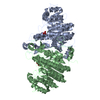
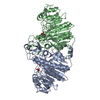
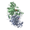
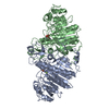
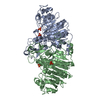
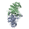
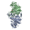
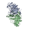
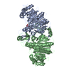
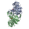
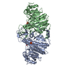
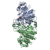
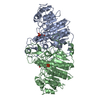
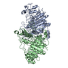
| Entry | Database: PDB / ID: 1khn | ||||||
|---|---|---|---|---|---|---|---|
| Title | E. COLI ALKALINE PHOSPHATASE MUTANT (D153HD330N) ZINC FORM | ||||||
 Components Components | Alkaline phosphatase | ||||||
 Keywords Keywords | HYDROLASE / ALKALINE PHOSPHATASE | ||||||
| Function / homology |  Function and homology information Function and homology informationoxidoreductase activity, acting on phosphorus or arsenic in donors / alkaline phosphatase / alkaline phosphatase activity / hydrogenase (acceptor) activity / phosphoprotein phosphatase activity / outer membrane-bounded periplasmic space / periplasmic space / magnesium ion binding / zinc ion binding Similarity search - Function | ||||||
| Biological species |  | ||||||
| Method |  X-RAY DIFFRACTION / SYNCHROTRON / FOURIER SYNTHESIS / Resolution: 2.6 Å X-RAY DIFFRACTION / SYNCHROTRON / FOURIER SYNTHESIS / Resolution: 2.6 Å | ||||||
 Authors Authors | Le Du, M.H. / Lamoure, C. / Muller, B.H. / Bulgakov, O.V. / Lajeunesse, E. / Menez, A. / Boulain, J.C. | ||||||
 Citation Citation | Journal: J.Mol.Biol. / Year: 2002 Title: Artificial evolution of an enzyme active site: structural studies of three highly active mutants of Escherichia coli alkaline phosphatase. Authors: Le Du, M.H. / Lamoure, C. / Muller, B.H. / Bulgakov, O.V. / Lajeunesse, E. / Menez, A. / Boulain, J.C. #1: Journal: J.Mol.Biol. / Year: 1991Title: Reaction Mechanism of Alkaline Phosphatase Based on Crystal Structures. Two-Metal Ion Catalysis Authors: Kim, E.E. / Wyckoff, H.W. | ||||||
| History |
|
- Structure visualization
| Structure viewer | Molecule: MolmilJmol/JSmol |
|---|
- Downloads & links
-Download
| PDBx/mmCIF format | 1khn.cif.gz | 179.6 KB | Display | PDBx/mmCIF format |
|---|---|---|---|---|
| PDB format | pdb1khn.ent.gz | 142.5 KB | Display | PDB format |
| PDBx/mmJSON format | 1khn.json.gz | Tree view | PDBx/mmJSON format | |
| Others |  Other downloads Other downloads |
-Validation report
| Arichive directory | https://data.pdbj.org/pub/pdb/validation_reports/kh/1khnftp://data.pdbj.org/pub/pdb/validation_reports/kh/1khn | HTTPS FTP |
|---|
-Related structure data
| Related structure data | 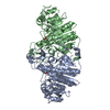 1kh4SC  1kh5C 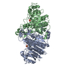 1kh7C  1kh9C 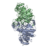 1khjC  1khkC  1khlC S: Starting model for refinement C: citing same article ( |
|---|---|
| Similar structure data |


-Links
 PDBj
PDBj
- Assembly
| Deposited unit | 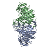
| ||||||||
|---|---|---|---|---|---|---|---|---|---|
| 1 |
| ||||||||
| Unit cell |
| ||||||||
| Noncrystallographic symmetry (NCS) | NCS oper: (Code: given Matrix: (0.00713, 0.999974, -0.001354), Vector: |
-Components
| #1: Protein | Mass: 47115.484 Da / Num. of mol.: 2 / Mutation: D153H, D330N Source method: isolated from a genetically manipulated source Source: (gene. exp.) #2: Chemical | ChemComp-ZN /   Mass: 65.409 Da / Num. of mol.: 6 / Source method: obtained synthetically / Formula: Zn Mass: 65.409 Da / Num. of mol.: 6 / Source method: obtained synthetically / Formula: Zn#3: Water | ChemComp-HOH / |  Mass: 18.015 Da / Num. of mol.: 311 / Source method: isolated from a natural source / Formula: H2O Mass: 18.015 Da / Num. of mol.: 311 / Source method: isolated from a natural source / Formula: H2OHas protein modification | Y | |
|---|
-Experimental details
-Experiment
| Experiment | Method: X-RAY DIFFRACTION / Number of used crystals: 1 |
|---|
- Sample preparation
| Crystal | Density Matthews: 2.82 Å3/Da / Density % sol: 56.46 % | ||||||||||||||||||||||||||||||||||||||||||
|---|---|---|---|---|---|---|---|---|---|---|---|---|---|---|---|---|---|---|---|---|---|---|---|---|---|---|---|---|---|---|---|---|---|---|---|---|---|---|---|---|---|---|---|
| Crystal grow | Temperature: 292 K / Method: vapor diffusion, hanging drop / pH: 8 Details: ammonium sulfate, magnesium chloride, zinc sulfate, TRIS, pH 8.0, VAPOR DIFFUSION, HANGING DROP, temperature 292K | ||||||||||||||||||||||||||||||||||||||||||
| Crystal grow | *PLUS Method: vapor diffusion | ||||||||||||||||||||||||||||||||||||||||||
| Components of the solutions | *PLUS
|
-Data collection
| Diffraction | Mean temperature: 292 K | |||||||||
|---|---|---|---|---|---|---|---|---|---|---|
| Diffraction source | Source: SYNCHROTRON / Site: ESRF  / Beamline: BM02 / Wavelength: 1.54 / Wavelength: 0.98 Å / Beamline: BM02 / Wavelength: 1.54 / Wavelength: 0.98 Å | |||||||||
| Detector | Type: MARRESEARCH / Detector: CCD / Date: Jul 1, 1996 | |||||||||
| Radiation | Monochromator: Mirror / Protocol: SINGLE WAVELENGTH / Monochromatic (M) / Laue (L): M / Scattering type: x-ray | |||||||||
| Radiation wavelength |
| |||||||||
| Reflection | Resolution: 2.6→20 Å / Num. all: 53663 / Num. obs: 53663 / % possible obs: 99.3 % / Observed criterion σ(F): 2 / Observed criterion σ(I): 0 / Rsym value: 0.0904 | |||||||||
| Reflection shell | Resolution: 2.6→2.74 Å / Rsym value: 0.46 / % possible all: 92 | |||||||||
| Reflection | *PLUS % possible obs: 96.3 % / Rmerge(I) obs: 0.0904 | |||||||||
| Reflection shell | *PLUS Rmerge(I) obs: 0.46 |
- Processing
| Software |
| |||||||||||||||||||||||||
|---|---|---|---|---|---|---|---|---|---|---|---|---|---|---|---|---|---|---|---|---|---|---|---|---|---|---|
| Refinement | Method to determine structure: FOURIER SYNTHESIS Starting model: PDB ENTRY 1KH4 Resolution: 2.6→10 Å / Cross valid method: IMPLOR-CYCLING TEST SETS / σ(F): 0 / Stereochemistry target values: XPLOR
| |||||||||||||||||||||||||
| Refinement step | Cycle: LAST / Resolution: 2.6→10 Å
| |||||||||||||||||||||||||
| LS refinement shell | Resolution: 2.6→2.74 Å / Total num. of bins used: 20
| |||||||||||||||||||||||||
| Refinement | *PLUS Highest resolution: 2.6 Å / Lowest resolution: 10 Å / σ(F): 0 / % reflection Rfree: 5 % | |||||||||||||||||||||||||
| Solvent computation | *PLUS | |||||||||||||||||||||||||
| Displacement parameters | *PLUS | |||||||||||||||||||||||||
| Refine LS restraints | *PLUS
| |||||||||||||||||||||||||
| LS refinement shell | *PLUS Highest resolution: 2.6 Å / % reflection Rfree: 5 % / Rfactor Rwork: 0.472 / Rfactor obs: 0.472 |
