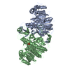
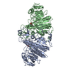
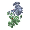
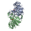
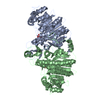
 Movie
Movie Controller


+ Open data

- Basic information
| Entry | Database: PDB / ID: 1hjk | ||||||
|---|---|---|---|---|---|---|---|
| Title | ALKALINE PHOSPHATASE MUTANT H331Q | ||||||
 Components Components | ALKALINE PHOSPHATASE | ||||||
 Keywords Keywords | HYDROLASE / ALKALINE PHOSPHATASE / PHOSPHORIC MONOESTER / TRANSFERASE(PHOSPHO / ALCOHOL ACCEPTOR) | ||||||
| Function / homology |  Function and homology information Function and homology informationoxidoreductase activity, acting on phosphorus or arsenic in donors / alkaline phosphatase / alkaline phosphatase activity / hydrogenase (acceptor) activity / phosphoprotein phosphatase activity / outer membrane-bounded periplasmic space / periplasmic space / magnesium ion binding / zinc ion binding Similarity search - Function | ||||||
| Biological species |  | ||||||
| Method |  X-RAY DIFFRACTION / Resolution: 2.3 Å X-RAY DIFFRACTION / Resolution: 2.3 Å | ||||||
 Authors Authors | Murphy, J.E. / Stec, B. / Ma, L. / Kantrowitz, E.R. | ||||||
 Citation Citation | Journal: Nat.Struct.Biol. / Year: 1997 Title: Trapping and visualization of a covalent enzyme-phosphate intermediate. Authors: Murphy, J.E. / Stec, B. / Ma, L. / Kantrowitz, E.R. #1: Journal: J.Mol.Biol. / Year: 1995Title: Mutations at Positions 153 and 328 in Escherichia Coli Alkaline Phosphatase Provide Insight Towards the Structure and Function of Mammalian and Yeast Alkaline Phosphatases Authors: Murphy, J.E. / Tibbitts, T.T. / Kantrowitz, E.R. #2: Journal: Protein Sci. / Year: 1994Title: Kinetics and Crystal Structure of a Mutant Escherichia Coli Alkaline Phosphatase (Asp-369-->Asn): A Mechanism Involving One Zinc Per Active Site Authors: Tibbitts, T.T. / Xu, X. / Kantrowitz, E.R. #3: Journal: J.Mol.Biol. / Year: 1991Title: Reaction Mechanism of Alkaline Phosphatase Based on Crystal Structures. Two-Metal Ion Catalysis Authors: Kim, E.E. / Wyckoff, H.W. | ||||||
| History |
|
- Structure visualization
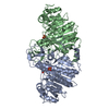
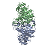
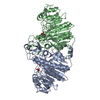
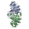
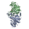
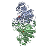
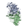
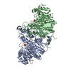
| Structure viewer | Molecule: MolmilJmol/JSmol |
|---|
- Downloads & links
-Download
| PDBx/mmCIF format | 1hjk.cif.gz | 183.8 KB | Display | PDBx/mmCIF format |
|---|---|---|---|---|
| PDB format | pdb1hjk.ent.gz | 145.6 KB | Display | PDB format |
| PDBx/mmJSON format | 1hjk.json.gz | Tree view | PDBx/mmJSON format | |
| Others |  Other downloads Other downloads |
-Validation report
| Arichive directory | https://data.pdbj.org/pub/pdb/validation_reports/hj/1hjkftp://data.pdbj.org/pub/pdb/validation_reports/hj/1hjk | HTTPS FTP |
|---|
-Related structure data
| Similar structure data |
|---|
-Links
 PDBj
PDBj
- Assembly
| Deposited unit | 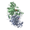
| |||||||||
|---|---|---|---|---|---|---|---|---|---|---|
| 1 |
| |||||||||
| Unit cell |
| |||||||||
| Components on special symmetry positions |
| |||||||||
| Noncrystallographic symmetry (NCS) | NCS domain:
|
-Components
| #1: Protein | Mass: 47164.359 Da / Num. of mol.: 2 / Mutation: H331Q Source method: isolated from a genetically manipulated source Source: (gene. exp.) #2: Chemical | ChemComp-ZN /   Mass: 65.409 Da / Num. of mol.: 4 / Source method: obtained synthetically / Formula: Zn Mass: 65.409 Da / Num. of mol.: 4 / Source method: obtained synthetically / Formula: Zn#3: Chemical |   Mass: 24.305 Da / Num. of mol.: 2 / Source method: obtained synthetically / Formula: Mg Mass: 24.305 Da / Num. of mol.: 2 / Source method: obtained synthetically / Formula: Mg#4: Chemical |   Mass: 96.063 Da / Num. of mol.: 2 / Source method: obtained synthetically / Formula: SO4 Mass: 96.063 Da / Num. of mol.: 2 / Source method: obtained synthetically / Formula: SO4#5: Water | ChemComp-HOH / |  Mass: 18.015 Da / Num. of mol.: 514 / Source method: isolated from a natural source / Formula: H2O Mass: 18.015 Da / Num. of mol.: 514 / Source method: isolated from a natural source / Formula: H2OHas protein modification | Y | |
|---|
-Experimental details
-Experiment
| Experiment | Method: X-RAY DIFFRACTION / Number of used crystals: 1 |
|---|
- Sample preparation
| Crystal | Density Matthews: 3.35 Å3/Da / Density % sol: 63.27 % |
|---|---|
| Crystal grow | pH: 7.5 Details: 55% SATURATING (NH4)2SO4, 100 MM TRIS, 10 MM MGCL2, 10 MM ZNCL2, 2 MM NAH2PO4 AT PH 7.5 |
| Crystal grow | *PLUS pH: 5.5 / Method: unknown |
| Components of the solutions | *PLUS Conc.: 0.1 M / Common name: sodium acetate |
-Data collection
| Diffraction | Mean temperature: 295 K |
|---|---|
| Diffraction source | Source: ROTATING ANODE / Type: RIGAKU RUH2R / Wavelength: 1.5418 |
| Detector | Type: XUONG-HAMLIN MULTIWIRE / Detector: AREA DETECTOR / Date: Nov 1, 1995 |
| Radiation | Monochromator: Y / Monochromatic (M) / Laue (L): M / Scattering type: x-ray |
| Radiation wavelength | Wavelength: 1.5418 Å / Relative weight: 1 |
| Reflection | Resolution: 2.3→80 Å / Num. obs: 51270 / % possible obs: 90 % / Observed criterion σ(I): 1 / Redundancy: 2.4 % / Rmerge(I) obs: 0.09 |
| Reflection | *PLUS Num. measured all: 120731 / Rmerge(I) obs: 0.09 |
- Processing
| Software |
| ||||||||||||||||||||||||||||||||||||||||||||||||||||||||||||
|---|---|---|---|---|---|---|---|---|---|---|---|---|---|---|---|---|---|---|---|---|---|---|---|---|---|---|---|---|---|---|---|---|---|---|---|---|---|---|---|---|---|---|---|---|---|---|---|---|---|---|---|---|---|---|---|---|---|---|---|---|---|
| Refinement | Resolution: 2.3→8 Å / σ(F): 2
| ||||||||||||||||||||||||||||||||||||||||||||||||||||||||||||
| Displacement parameters | Biso mean: 13.15 Å2 | ||||||||||||||||||||||||||||||||||||||||||||||||||||||||||||
| Refine analyze |
| ||||||||||||||||||||||||||||||||||||||||||||||||||||||||||||
| Refinement step | Cycle: LAST / Resolution: 2.3→8 Å
| ||||||||||||||||||||||||||||||||||||||||||||||||||||||||||||
| Refine LS restraints |
| ||||||||||||||||||||||||||||||||||||||||||||||||||||||||||||
| Refine LS restraints NCS |
| ||||||||||||||||||||||||||||||||||||||||||||||||||||||||||||
| Software | *PLUS Name: X-PLOR / Classification: refinement | ||||||||||||||||||||||||||||||||||||||||||||||||||||||||||||
| Refinement | *PLUS Rfactor Rfree: 0.2 | ||||||||||||||||||||||||||||||||||||||||||||||||||||||||||||
| Solvent computation | *PLUS | ||||||||||||||||||||||||||||||||||||||||||||||||||||||||||||
| Displacement parameters | *PLUS | ||||||||||||||||||||||||||||||||||||||||||||||||||||||||||||
| Refine LS restraints | *PLUS
|
