 Movie
Movie Controller


+ Open data

- Basic information
| Entry | Database: PDB / ID: 1alj | ||||||
|---|---|---|---|---|---|---|---|
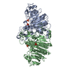
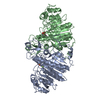
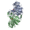
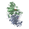
| Title | ALKALINE PHOSPHATASE MUTANT (H412N) | ||||||
 Components Components | ALKALINE PHOSPHATASE | ||||||
 Keywords Keywords | HYDROLASE (PHOSPHORIC MONOESTER) / TRANSFERASE (PHOSPHO / ALCOHOL ACCEPTOR) | ||||||
| Function / homology |  Function and homology information Function and homology informationoxidoreductase activity, acting on phosphorus or arsenic in donors / alkaline phosphatase / alkaline phosphatase activity / hydrogenase (acceptor) activity / dephosphorylation / phosphoprotein phosphatase activity / outer membrane-bounded periplasmic space / periplasmic space / magnesium ion binding / zinc ion binding Similarity search - Function | ||||||
| Biological species |  | ||||||
| Method |  X-RAY DIFFRACTION / Resolution: 2.6 Å X-RAY DIFFRACTION / Resolution: 2.6 Å | ||||||
 Authors Authors | Ma, L. / Tibbitts, T.T. / Kantrowitz, E.R. | ||||||
 Citation Citation | Journal: Protein Sci. / Year: 1995 Title: Escherichia coli alkaline phosphatase: X-ray structural studies of a mutant enzyme (His-412-->Asn) at one of the catalytically important zinc binding sites. Authors: Ma, L. / Tibbitts, T.T. / Kantrowitz, E.R. #1: Journal: J.Mol.Biol. / Year: 1991Title: Reaction Mechanism of Alkaline Phosphatase Based on Crystal Structures. Two Metal Ion Catalysis Authors: Kim, E.E. / Wyckoff, H.W. #2: Journal: J.Biol.Chem. / Year: 1994Title: Mutations at Histidine 412 Alter Zinc Binding and Eliminate Transferase Activity in Escherichia Coli Alkaline Phosphatase Authors: Ma, L. / Kantrowitz, E.R. | ||||||
| History |
|
- Structure visualization
| Structure viewer | Molecule: MolmilJmol/JSmol |
|---|
- Downloads & links
-Download
| PDBx/mmCIF format | 1alj.cif.gz | 176 KB | Display | PDBx/mmCIF format |
|---|---|---|---|---|
| PDB format | pdb1alj.ent.gz | 139.7 KB | Display | PDB format |
| PDBx/mmJSON format | 1alj.json.gz | Tree view | PDBx/mmJSON format | |
| Others |  Other downloads Other downloads |
-Validation report
| Arichive directory | https://data.pdbj.org/pub/pdb/validation_reports/al/1aljftp://data.pdbj.org/pub/pdb/validation_reports/al/1alj | HTTPS FTP |
|---|
-Related structure data


-Links
 PDBj
PDBj
- Assembly
| Deposited unit | 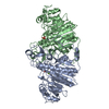
| ||||||||
|---|---|---|---|---|---|---|---|---|---|
| 1 |
| ||||||||
| Unit cell |
| ||||||||
| Details | THERE IS A DIMER (IDENTICAL CHAINS OF 449 RESIDUES) PER ASYMMETRIC UNIT. THESE SUBUNITS, DESIGNATED "A" AND "B". THE FIRST THREE RESIDUES WERE DELETED DURING THE REFINEMENT. |
-Components
| #1: Protein | Mass: 47070.355 Da / Num. of mol.: 2 / Mutation: H412N Source method: isolated from a genetically manipulated source Source: (gene. exp.) #2: Chemical |   Mass: 65.409 Da / Num. of mol.: 2 / Source method: obtained synthetically / Formula: Zn Mass: 65.409 Da / Num. of mol.: 2 / Source method: obtained synthetically / Formula: Zn#3: Chemical |   Mass: 24.305 Da / Num. of mol.: 2 / Source method: obtained synthetically / Formula: Mg Mass: 24.305 Da / Num. of mol.: 2 / Source method: obtained synthetically / Formula: Mg#4: Chemical |   Mass: 94.971 Da / Num. of mol.: 2 / Source method: obtained synthetically / Formula: PO4 Mass: 94.971 Da / Num. of mol.: 2 / Source method: obtained synthetically / Formula: PO4#5: Water | ChemComp-HOH / |  Mass: 18.015 Da / Num. of mol.: 219 / Source method: isolated from a natural source / Formula: H2O Mass: 18.015 Da / Num. of mol.: 219 / Source method: isolated from a natural source / Formula: H2OHas protein modification | Y | |
|---|
-Experimental details
-Experiment
| Experiment | Method: X-RAY DIFFRACTION |
|---|
- Sample preparation
| Crystal | Density Matthews: 3.31 Å3/Da / Density % sol: 62.79 % | ||||||||||||||||||||||||
|---|---|---|---|---|---|---|---|---|---|---|---|---|---|---|---|---|---|---|---|---|---|---|---|---|---|
| Crystal grow | Details: THIS ENTRY IS A MUTANT ALKALINE PHOSPHATE (H412N) IN WHICH HIS 412 IS REPLACED BY ASN, DETERMINED WITH CRYSTALS SOAKED IN STABILIZATION BUFFER CONTAINING NO ADDED ZINC. THERE IS ONE ZINC AND ...Details: THIS ENTRY IS A MUTANT ALKALINE PHOSPHATE (H412N) IN WHICH HIS 412 IS REPLACED BY ASN, DETERMINED WITH CRYSTALS SOAKED IN STABILIZATION BUFFER CONTAINING NO ADDED ZINC. THERE IS ONE ZINC AND ONE MAGNESIUM COMPLEXED WITH INORGANIC PHOSPHATE BOUND IN EACH OF THE TWO ACTIVE SITES. | ||||||||||||||||||||||||
| Crystal grow | *PLUS pH: 9.5 / Method: vapor diffusion, hanging drop | ||||||||||||||||||||||||
| Components of the solutions | *PLUS
|
-Data collection
| Diffraction source | Wavelength: 1.54 |
|---|---|
| Detector | Type: ADSC / Detector: AREA DETECTOR / Date: May 14, 1993 |
| Radiation | Monochromatic (M) / Laue (L): M / Scattering type: x-ray |
| Radiation wavelength | Wavelength: 1.54 Å / Relative weight: 1 |
| Reflection | Resolution: 2.5→26.3 Å / Num. obs: 36882 / % possible obs: 98.4 % / Observed criterion σ(I): 3 / Redundancy: 4.6 % / Rmerge(I) obs: 0.108 |
| Reflection | *PLUS Rmerge(I) obs: 0.108 |
- Processing
| Software |
| ||||||||||||||||||||||||||||||||||||||||||||||||||||||||||||
|---|---|---|---|---|---|---|---|---|---|---|---|---|---|---|---|---|---|---|---|---|---|---|---|---|---|---|---|---|---|---|---|---|---|---|---|---|---|---|---|---|---|---|---|---|---|---|---|---|---|---|---|---|---|---|---|---|---|---|---|---|---|
| Refinement | Resolution: 2.6→8 Å / σ(F): 0 /
| ||||||||||||||||||||||||||||||||||||||||||||||||||||||||||||
| Refine analyze | Luzzati coordinate error obs: 0.27 Å | ||||||||||||||||||||||||||||||||||||||||||||||||||||||||||||
| Refinement step | Cycle: LAST / Resolution: 2.6→8 Å
| ||||||||||||||||||||||||||||||||||||||||||||||||||||||||||||
| Refine LS restraints |
| ||||||||||||||||||||||||||||||||||||||||||||||||||||||||||||
| Software | *PLUS Name: X-PLOR / Classification: refinement | ||||||||||||||||||||||||||||||||||||||||||||||||||||||||||||
| Refinement | *PLUS | ||||||||||||||||||||||||||||||||||||||||||||||||||||||||||||
| Solvent computation | *PLUS | ||||||||||||||||||||||||||||||||||||||||||||||||||||||||||||
| Displacement parameters | *PLUS | ||||||||||||||||||||||||||||||||||||||||||||||||||||||||||||
| Refine LS restraints | *PLUS
|
