 Movie
Movie Controller


+ Open data

- Basic information
| Entry | Database: PDB / ID: 1ani | ||||||
|---|---|---|---|---|---|---|---|
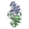
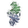
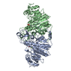
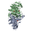
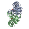
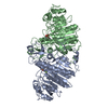
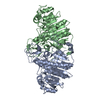
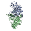
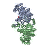
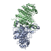
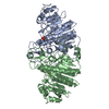
| Title | ALKALINE PHOSPHATASE (D153H, K328H) | ||||||
 Components Components | ALKALINE PHOSPHATASE | ||||||
 Keywords Keywords | ALKALINE PHOSPHATASE / HYDROLASE (PHOSPHORIC MONOESTER) / TRANSFERASE (PHOSPHO / ALCOHOL ACCEPTOR) | ||||||
| Function / homology |  Function and homology information Function and homology informationoxidoreductase activity, acting on phosphorus or arsenic in donors / alkaline phosphatase / alkaline phosphatase activity / hydrogenase (acceptor) activity / dephosphorylation / phosphoprotein phosphatase activity / outer membrane-bounded periplasmic space / periplasmic space / magnesium ion binding / zinc ion binding Similarity search - Function | ||||||
| Biological species |  | ||||||
| Method |  X-RAY DIFFRACTION / Resolution: 2.5 Å X-RAY DIFFRACTION / Resolution: 2.5 Å | ||||||
 Authors Authors | Murphy, J.E. / Tibbitts, T.T. / Kantrowitz, E.R. | ||||||
 Citation Citation | Journal: J.Mol.Biol. / Year: 1995 Title: Mutations at positions 153 and 328 in Escherichia coli alkaline phosphatase provide insight towards the structure and function of mammalian and yeast alkaline phosphatases. Authors: Murphy, J.E. / Tibbitts, T.T. / Kantrowitz, E.R. #1: Journal: Biochemistry / Year: 1993Title: Magnesium in the Active Site of Escherichia Coli Alkaline Phosphatase is Important for Both Structural Stabilization and Catalysis Authors: Janeway, C.M.L. / Xu, X. / Murphy, J.E. / Chaidaroglou, A. / Kantrowitz, E.R. #2: Journal: J.Biol.Chem. / Year: 1993Title: Conversion of a Magnesium Binding Site Into a Zinc Binding Site by a Single Amino Acid Substitution in Escherichia Coli Alkaline Phosphatase Authors: Murphy, J.E. / Xu, X. / Kantrowitz, E.R. #3: Journal: J.Mol.Biol. / Year: 1991Title: Reaction Mechanism of Alkaline Phosphatase Based on Crystal Structures. Two Metal Ion Catalysis Authors: Kim, E.E. / Wyckoff, H.W. | ||||||
| History |
|
- Structure visualization
| Structure viewer | Molecule: MolmilJmol/JSmol |
|---|
- Downloads & links
-Download
| PDBx/mmCIF format | 1ani.cif.gz | 178.9 KB | Display | PDBx/mmCIF format |
|---|---|---|---|---|
| PDB format | pdb1ani.ent.gz | 140.8 KB | Display | PDB format |
| PDBx/mmJSON format | 1ani.json.gz | Tree view | PDBx/mmJSON format | |
| Others |  Other downloads Other downloads |
-Validation report
| Arichive directory | https://data.pdbj.org/pub/pdb/validation_reports/an/1aniftp://data.pdbj.org/pub/pdb/validation_reports/an/1ani | HTTPS FTP |
|---|
-Related structure data


-Links
 PDBj
PDBj
- Assembly
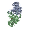
| Deposited unit | 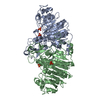
| ||||||||
|---|---|---|---|---|---|---|---|---|---|
| 1 |
| ||||||||
| Unit cell |
|
-Components
| #1: Protein | Mass: 46799.086 Da / Num. of mol.: 2 / Mutation: D153H, K328H Source method: isolated from a genetically manipulated source Details: 65% SATURATING (NH4)2SO4, 100 MM TRIS, 10 MM MGCL2, 10 MM ZNCL2, 2 MM NAH2PO4 AT PH 7.5 Source: (gene. exp.) #2: Chemical | ChemComp-ZN /   Mass: 65.409 Da / Num. of mol.: 6 / Source method: obtained synthetically / Formula: Zn Mass: 65.409 Da / Num. of mol.: 6 / Source method: obtained synthetically / Formula: Zn#3: Chemical | ChemComp-PO4 /   Mass: 94.971 Da / Num. of mol.: 5 / Source method: obtained synthetically / Formula: PO4 Mass: 94.971 Da / Num. of mol.: 5 / Source method: obtained synthetically / Formula: PO4#4: Water | ChemComp-HOH / |  Mass: 18.015 Da / Num. of mol.: 270 / Source method: isolated from a natural source / Formula: H2O Mass: 18.015 Da / Num. of mol.: 270 / Source method: isolated from a natural source / Formula: H2OHas protein modification | Y | |
|---|
-Experimental details
-Experiment
| Experiment | Method: X-RAY DIFFRACTION |
|---|
- Sample preparation
| Crystal | Density Matthews: 3.32 Å3/Da / Density % sol: 62.91 % | ||||||||||||||||||||||||||||||||||||||||||
|---|---|---|---|---|---|---|---|---|---|---|---|---|---|---|---|---|---|---|---|---|---|---|---|---|---|---|---|---|---|---|---|---|---|---|---|---|---|---|---|---|---|---|---|
| Crystal grow | pH: 7.5 / Details: pH 7.5 | ||||||||||||||||||||||||||||||||||||||||||
| Crystal grow | *PLUS pH: 9.5 / Method: vapor diffusion, hanging drop | ||||||||||||||||||||||||||||||||||||||||||
| Components of the solutions | *PLUS
|
-Data collection
| Diffraction source | Wavelength: 1.5418 Å |
|---|---|
| Detector | Type: SAN DIEGO MULTIWIRE / Detector: AREA DETECTOR / Date: Oct 28, 1993 |
| Radiation | Monochromatic (M) / Laue (L): M / Scattering type: x-ray |
| Radiation wavelength | Wavelength: 1.5418 Å / Relative weight: 1 |
| Reflection | Resolution: 2.5→8 Å / Num. obs: 39775 / % possible obs: 91 % / Observed criterion σ(I): 3 / Redundancy: 3 % / Rmerge(I) obs: 0.072 |
| Reflection | *PLUS Num. measured all: 118201 / Rmerge(I) obs: 0.072 |
- Processing
| Software |
| ||||||||||||||||||||||||||||||||||||||||||||||||||||||||||||
|---|---|---|---|---|---|---|---|---|---|---|---|---|---|---|---|---|---|---|---|---|---|---|---|---|---|---|---|---|---|---|---|---|---|---|---|---|---|---|---|---|---|---|---|---|---|---|---|---|---|---|---|---|---|---|---|---|---|---|---|---|---|
| Refinement | Resolution: 2.5→8 Å / σ(F): 2
| ||||||||||||||||||||||||||||||||||||||||||||||||||||||||||||
| Refine analyze | Luzzati coordinate error obs: 0.25 Å | ||||||||||||||||||||||||||||||||||||||||||||||||||||||||||||
| Refinement step | Cycle: LAST / Resolution: 2.5→8 Å
| ||||||||||||||||||||||||||||||||||||||||||||||||||||||||||||
| Refine LS restraints |
| ||||||||||||||||||||||||||||||||||||||||||||||||||||||||||||
| Refinement | *PLUS Rfactor Rfree: 0.233 | ||||||||||||||||||||||||||||||||||||||||||||||||||||||||||||
| Solvent computation | *PLUS | ||||||||||||||||||||||||||||||||||||||||||||||||||||||||||||
| Displacement parameters | *PLUS | ||||||||||||||||||||||||||||||||||||||||||||||||||||||||||||
| Refine LS restraints | *PLUS
|
