 Movie
Movie Controller

[English] 日本語
 Yorodumi
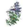
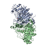
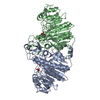
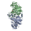
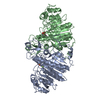
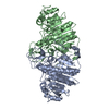
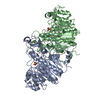
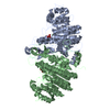
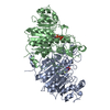
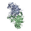
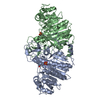
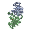
- PDB-1ew8: ALKALINE PHOSPHATASE (E.C. 3.1.3.1) COMPLEX WITH PHOSPHONOACETIC ACID -
+ Open data

- Basic information
| Entry | Database: PDB / ID: 1ew8 | ||||||
|---|---|---|---|---|---|---|---|
| Title | ALKALINE PHOSPHATASE (E.C. 3.1.3.1) COMPLEX WITH PHOSPHONOACETIC ACID | ||||||
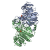
 Components Components | ALKALINE PHOSPHATASE | ||||||
 Keywords Keywords | HYDROLASE / enzyme-inhibitor complex | ||||||
| Function / homology |  Function and homology information Function and homology informationoxidoreductase activity, acting on phosphorus or arsenic in donors / alkaline phosphatase / alkaline phosphatase activity / hydrogenase (acceptor) activity / dephosphorylation / phosphoprotein phosphatase activity / outer membrane-bounded periplasmic space / periplasmic space / magnesium ion binding / zinc ion binding Similarity search - Function | ||||||
| Biological species |  | ||||||
| Method |  X-RAY DIFFRACTION / Resolution: 2.2 Å X-RAY DIFFRACTION / Resolution: 2.2 Å | ||||||
 Authors Authors | Holtz, K.M. / Stec, B. / Myers, J.K. / Antonelli, S.M. / Widlanski, T.S. / Kantrowitz, E.R. | ||||||
 Citation Citation | Journal: Protein Sci. / Year: 2000 Title: Alternate modes of binding in two crystal structures of alkaline phosphatase-inhibitor complexes. Authors: Holtz, K.M. / Stec, B. / Myers, J.K. / Antonelli, S.M. / Widlanski, T.S. / Kantrowitz, E.R. #1: Journal: J.Biol.Chem. / Year: 1999Title: A Model of the Transition State in the Alkaline Phosphatase Reaction Authors: Holtz, K.M. / Stec, B. / Kantrowitz, E.R. #2: Journal: J.Mol.Biol. / Year: 1991Title: Reaction Mechanism of Alkaline Phosphatase Based on Crystal Structures: Two-metal Ion Catalysis Authors: Kim, E.E. / Wyckoff, H.W. | ||||||
| History |
|
- Structure visualization
| Structure viewer | Molecule: MolmilJmol/JSmol |
|---|
- Downloads & links
-Download
| PDBx/mmCIF format | 1ew8.cif.gz | 189.3 KB | Display | PDBx/mmCIF format |
|---|---|---|---|---|
| PDB format | pdb1ew8.ent.gz | 149 KB | Display | PDB format |
| PDBx/mmJSON format | 1ew8.json.gz | Tree view | PDBx/mmJSON format | |
| Others |  Other downloads Other downloads |
-Validation report
| Arichive directory | https://data.pdbj.org/pub/pdb/validation_reports/ew/1ew8ftp://data.pdbj.org/pub/pdb/validation_reports/ew/1ew8 | HTTPS FTP |
|---|
-Related structure data


-Links
 PDBj
PDBj
- Assembly
| Deposited unit | 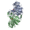
| ||||||||
|---|---|---|---|---|---|---|---|---|---|
| 1 |
| ||||||||
| Unit cell |
| ||||||||
| Details | homodimeric metalloenzyme with a non-crystallographic 2-fold symmetry axis |
-Components
-Protein , 1 types, 2 molecules AB
| #1: Protein | Mass: 47094.398 Da / Num. of mol.: 2 Source method: isolated from a genetically manipulated source Source: (gene. exp.) |
|---|
-Non-polymers , 6 types, 531 molecules 


| #2: Chemical | ChemComp-ZN /  Mass: 65.409 Da / Num. of mol.: 6 / Source method: obtained synthetically / Formula: Zn / Details: Obtained from Sigma Chemical Co. Mass: 65.409 Da / Num. of mol.: 6 / Source method: obtained synthetically / Formula: Zn / Details: Obtained from Sigma Chemical Co.#3: Chemical |  Mass: 94.971 Da / Num. of mol.: 2 / Source method: obtained synthetically / Formula: PO4 Mass: 94.971 Da / Num. of mol.: 2 / Source method: obtained synthetically / Formula: PO4#4: Chemical |  Mass: 96.063 Da / Num. of mol.: 2 / Source method: obtained synthetically / Formula: SO4 Mass: 96.063 Da / Num. of mol.: 2 / Source method: obtained synthetically / Formula: SO4#5: Chemical |  Mass: 24.305 Da / Num. of mol.: 2 / Source method: obtained synthetically / Formula: Mg Mass: 24.305 Da / Num. of mol.: 2 / Source method: obtained synthetically / Formula: Mg#6: Chemical |  Mass: 140.032 Da / Num. of mol.: 2 / Source method: obtained synthetically / Formula: C2H5O5P Mass: 140.032 Da / Num. of mol.: 2 / Source method: obtained synthetically / Formula: C2H5O5P#7: Water | ChemComp-HOH / | Mass: 18.015 Da / Num. of mol.: 517 / Source method: isolated from a natural source / Formula: H2O |
|---|
-Details
| Has protein modification | Y |
|---|---|
| Nonpolymer details | The active site has mixed occupancy with some phosphonoacetic acid (PAE) and some phosphate (PO4). ...The active site has mixed occupancy with some phosphonoacetic acid (PAE) and some phosphate (PO4). The PAE and PO4 are assigned different residue numbers, 556 and 557, and alternate conformation indicators A and B, respectively. The alternate position indicator signifies that these residues occupy the same area in space. The magnesium 452 has three waters associated with it and it has been labelled as ligand MO3 for each chain. The zinc ion 452 has the same three waters associated with it and is labelled ZO3. The ZO3 and MO3 occupy the same space and have the same coordinates, but their occupancies are different. The MO3 is labelled as residue 452, conformation A. The ZO3 is labelled as residue 453, conformation B. SO4 558 is associated with chain A, and SO4 568 is associated with chain B. |
-Experimental details
-Experiment
| Experiment | Method: X-RAY DIFFRACTION / Number of used crystals: 1 |
|---|
- Sample preparation
| Crystal | Density Matthews: 3.31 Å3/Da / Density % sol: 62.82 % | ||||||||||||||||||||||||||||||||||||||||||
|---|---|---|---|---|---|---|---|---|---|---|---|---|---|---|---|---|---|---|---|---|---|---|---|---|---|---|---|---|---|---|---|---|---|---|---|---|---|---|---|---|---|---|---|
| Crystal grow | Temperature: 298 K / Method: vapor diffusion, hanging drop / pH: 9.5 Details: enzyme: 30 mg/mL; buffer: 40% saturating ammonium sulfate/100 mM Tris/10 mM MgSO4, pH 9.5, VAPOR DIFFUSION, HANGING DROP, temperature 298K | ||||||||||||||||||||||||||||||||||||||||||
| Crystal grow | *PLUS | ||||||||||||||||||||||||||||||||||||||||||
| Components of the solutions | *PLUS
|
-Data collection
| Diffraction | Mean temperature: 295 K |
|---|---|
| Diffraction source | Source: ROTATING ANODE / Type: RIGAKU RU200 / Wavelength: 1.5418 |
| Detector | Type: UCSD MARK III / Detector: AREA DETECTOR / Date: Jun 5, 1996 |
| Radiation | Protocol: SINGLE WAVELENGTH / Monochromatic (M) / Laue (L): M / Scattering type: x-ray |
| Radiation wavelength | Wavelength: 1.5418 Å / Relative weight: 1 |
| Reflection | Resolution: 2.2→30 Å / Num. all: 63777 / Num. obs: 59057 / % possible obs: 92.6 % / Observed criterion σ(F): 0 / Observed criterion σ(I): 0 / Redundancy: 3.1 % / Biso Wilson estimate: 31.9 Å2 / Rmerge(I) obs: 0.078 / Net I/σ(I): 6.6 |
| Reflection shell | Resolution: 2.2→2.37 Å / Redundancy: 1.92 % / Rmerge(I) obs: 0.28 / Num. unique all: 11823 / % possible all: 94 |
| Reflection | *PLUS Num. measured all: 180502 |
| Reflection shell | *PLUS Lowest resolution: 2.4 Å / % possible obs: 79.6 % / Redundancy: 2 % / Mean I/σ(I) obs: 1.2 |
- Processing
| Software |
| |||||||||||||||||||||||||
|---|---|---|---|---|---|---|---|---|---|---|---|---|---|---|---|---|---|---|---|---|---|---|---|---|---|---|
| Refinement | Resolution: 2.2→12 Å / σ(F): 0 / σ(I): 0 / Stereochemistry target values: Engh & Huber Details: Used conjugated gradient least squares for the refinement and weighted full matrix least squares procedure for estimating ESDs.
| |||||||||||||||||||||||||
| Refinement step | Cycle: LAST / Resolution: 2.2→12 Å
| |||||||||||||||||||||||||
| Refine LS restraints |
| |||||||||||||||||||||||||
| Software | *PLUS Name: SHELXL / Version: 97 / Classification: refinement | |||||||||||||||||||||||||
| Refinement | *PLUS % reflection Rfree: 10 % / Rfactor all: 0.19 / Rfactor Rwork: 0.19 | |||||||||||||||||||||||||
| Solvent computation | *PLUS | |||||||||||||||||||||||||
| Displacement parameters | *PLUS | |||||||||||||||||||||||||
| Refine LS restraints | *PLUS
|
