 Movie
Movie Controller

[English] 日本語
 Yorodumi
- PDB-1ajb: THREE-DIMENSIONAL STRUCTURE OF THE D153G MUTANT OF E. COLI ALKALI... -
+ Open data

- Basic information
| Entry | Database: PDB / ID: 1ajb | ||||||
|---|---|---|---|---|---|---|---|
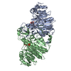
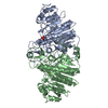
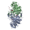
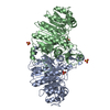
| Title | THREE-DIMENSIONAL STRUCTURE OF THE D153G MUTANT OF E. COLI ALKALINE PHOSPHATASE: A MUTANT WITH WEAKER MAGNESIUM BINDING AND INCREASED CATALYTIC ACTIVITY | ||||||
 Components Components | ALKALINE PHOSPHATASE | ||||||
 Keywords Keywords | NON SPECIFIC MONO-ESTERASE | ||||||
| Function / homology |  Function and homology information Function and homology informationoxidoreductase activity, acting on phosphorus or arsenic in donors / alkaline phosphatase / alkaline phosphatase activity / hydrogenase (acceptor) activity / dephosphorylation / phosphoprotein phosphatase activity / outer membrane-bounded periplasmic space / periplasmic space / magnesium ion binding / zinc ion binding Similarity search - Function | ||||||
| Biological species |  | ||||||
| Method |  X-RAY DIFFRACTION / Resolution: 2.5 Å X-RAY DIFFRACTION / Resolution: 2.5 Å | ||||||
 Authors Authors | Dealwis, C.G. / Chen, L. / Abad-Zapatero, C. | ||||||
 Citation Citation | Journal: Protein Eng. / Year: 1995 Title: 3-D structure of the D153G mutant of Escherichia coli alkaline phosphatase: an enzyme with weaker magnesium binding and increased catalytic activity. Authors: Dealwis, C.G. / Chen, L. / Brennan, C. / Mandecki, W. / Abad-Zapatero, C. #1: Journal: To be PublishedTitle: Crystallographic Analysis of Reversible Metal Binding Observed in a Mutant (Asp 153--> Gly) of E. Coli Alkaline Phosphatase Authors: Dealwis, C. / Brennan, C. / Christianson, K. / Mandecki, W. / Abad-Zapatero, A. #2: Journal: Protein Eng. / Year: 1992Title: 3-D Structure of the (Asp 101-->Ser) of E.Coli Alkaline Phosphatase with Higher Catalytic Activity Authors: Chen, L. / Neidhart, D. / Kohlbrenner, M. / Mandecki, W. / Bell, S. / Sowadski, J. / Abad-Zapatero, C. | ||||||
| History |
| ||||||
| Remark 700 | SHEET SHEET DETERMINATION METHOD: KABSCH & SANDER; SHEET_ID: S1A; SAME AS 1ALK. SHEET_ID: S2A; SAME ...SHEET SHEET DETERMINATION METHOD: KABSCH & SANDER; SHEET_ID: S1A; SAME AS 1ALK. SHEET_ID: S2A; SAME AS 1ALK. SHEET_ID: S1B; SAME AS 1ALK. SHEET_ID: S2B; SAME AS 1ALK. |
- Structure visualization
| Structure viewer | Molecule: MolmilJmol/JSmol |
|---|
- Downloads & links
-Download
| PDBx/mmCIF format | 1ajb.cif.gz | 175.5 KB | Display | PDBx/mmCIF format |
|---|---|---|---|---|
| PDB format | pdb1ajb.ent.gz | 138.8 KB | Display | PDB format |
| PDBx/mmJSON format | 1ajb.json.gz | Tree view | PDBx/mmJSON format | |
| Others |  Other downloads Other downloads |
-Validation report
| Arichive directory | https://data.pdbj.org/pub/pdb/validation_reports/aj/1ajbftp://data.pdbj.org/pub/pdb/validation_reports/aj/1ajb | HTTPS FTP |
|---|
-Related structure data
| Similar structure data |
|---|
-Links
 PDBj
PDBj
- Assembly
| Deposited unit | 
| ||||||||
|---|---|---|---|---|---|---|---|---|---|
| 1 |
| ||||||||
| Unit cell |
|
-Components
| #1: Protein | Mass: 47037.348 Da / Num. of mol.: 2 / Mutation: D153G Source method: isolated from a genetically manipulated source Details: MUTANT WITH 4-FOLD INCREASED ACTIVITY AND WEAKER MG BINDING Source: (gene. exp.) Description: LAC PROMOTER. FOR MORE INFORMATION ABOUT THE EXPRESSION SYSTEM CONSULT MANDECKI ET AL. GENE 94, 103-107; (1990). Gene: PHOA / Plasmid: PWM528 AS BAMH1/HINDIII RESTRICTION FRAGMENT / Gene (production host): PHOA / Production host: #2: Chemical | ChemComp-ZN /   Mass: 65.409 Da / Num. of mol.: 4 / Source method: obtained synthetically / Formula: Zn Mass: 65.409 Da / Num. of mol.: 4 / Source method: obtained synthetically / Formula: Zn#3: Chemical |   Mass: 24.305 Da / Num. of mol.: 2 / Source method: obtained synthetically / Formula: Mg Mass: 24.305 Da / Num. of mol.: 2 / Source method: obtained synthetically / Formula: Mg#4: Chemical |   Mass: 96.063 Da / Num. of mol.: 2 / Source method: obtained synthetically / Formula: SO4 Mass: 96.063 Da / Num. of mol.: 2 / Source method: obtained synthetically / Formula: SO4#5: Water | ChemComp-HOH / |  Mass: 18.015 Da / Num. of mol.: 167 / Source method: isolated from a natural source / Formula: H2O Mass: 18.015 Da / Num. of mol.: 167 / Source method: isolated from a natural source / Formula: H2OHas protein modification | Y | Source details | FOR MORE INFORMATIO | |
|---|
-Experimental details
-Experiment
| Experiment | Method: X-RAY DIFFRACTION |
|---|
- Sample preparation
| Crystal | Density Matthews: 3.31 Å3/Da / Density % sol: 62.78 % | |||||||||||||||||||||||||
|---|---|---|---|---|---|---|---|---|---|---|---|---|---|---|---|---|---|---|---|---|---|---|---|---|---|---|
| Crystal grow | *PLUS pH: 9.5 / Method: vapor diffusion, hanging drop | |||||||||||||||||||||||||
| Components of the solutions | *PLUS
|
-Data collection
| Diffraction source | Wavelength: 1.5418 Å |
|---|---|
| Detector | Type: RIGAKU RAXIS / Detector: IMAGE PLATE / Date: Jan 6, 1994 |
| Radiation | Monochromatic (M) / Laue (L): M / Scattering type: x-ray |
| Radiation wavelength | Wavelength: 1.5418 Å / Relative weight: 1 |
| Reflection | Num. obs: 40175 / % possible obs: 77 % / Observed criterion σ(I): 0 / Redundancy: 1.75 % / Rmerge(I) obs: 0.077 |
| Reflection | *PLUS Highest resolution: 2.5 Å / Rmerge(I) obs: 0.077 |
- Processing
| Software |
| ||||||||||||||||||||||||||||||||||||||||||||||||||||||||||||
|---|---|---|---|---|---|---|---|---|---|---|---|---|---|---|---|---|---|---|---|---|---|---|---|---|---|---|---|---|---|---|---|---|---|---|---|---|---|---|---|---|---|---|---|---|---|---|---|---|---|---|---|---|---|---|---|---|---|---|---|---|---|
| Refinement | Resolution: 2.5→6 Å / σ(F): 0 /
| ||||||||||||||||||||||||||||||||||||||||||||||||||||||||||||
| Displacement parameters | Biso mean: 11 Å2 | ||||||||||||||||||||||||||||||||||||||||||||||||||||||||||||
| Refine analyze | Luzzati coordinate error obs: 0.2 Å | ||||||||||||||||||||||||||||||||||||||||||||||||||||||||||||
| Refinement step | Cycle: LAST / Resolution: 2.5→6 Å
| ||||||||||||||||||||||||||||||||||||||||||||||||||||||||||||
| Refine LS restraints |
| ||||||||||||||||||||||||||||||||||||||||||||||||||||||||||||
| Software | *PLUS Name: X-PLOR / Classification: refinement | ||||||||||||||||||||||||||||||||||||||||||||||||||||||||||||
| Refinement | *PLUS | ||||||||||||||||||||||||||||||||||||||||||||||||||||||||||||
| Solvent computation | *PLUS | ||||||||||||||||||||||||||||||||||||||||||||||||||||||||||||
| Displacement parameters | *PLUS | ||||||||||||||||||||||||||||||||||||||||||||||||||||||||||||
| Refine LS restraints | *PLUS
|
