 Movie
Movie Controller

[English] 日本語
 Yorodumi
- PDB-1y6v: Structure of E. coli Alkaline Phosphatase in presence of cobalt a... -
+ Open data

- Basic information
| Entry | Database: PDB / ID: 1y6v | ||||||
|---|---|---|---|---|---|---|---|
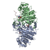
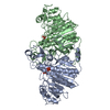
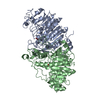
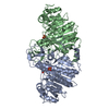
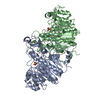
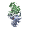
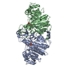
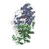
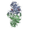
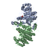
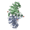
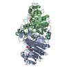
| Title | Structure of E. coli Alkaline Phosphatase in presence of cobalt at 1.60 A resolution | ||||||
 Components Components | Alkaline phosphatase | ||||||
 Keywords Keywords | HYDROLASE / metal specificity / high-spin/low-spin configurations | ||||||
| Function / homology |  Function and homology information Function and homology informationoxidoreductase activity, acting on phosphorus or arsenic in donors / alkaline phosphatase / alkaline phosphatase activity / hydrogenase (acceptor) activity / phosphoprotein phosphatase activity / outer membrane-bounded periplasmic space / periplasmic space / magnesium ion binding / zinc ion binding Similarity search - Function | ||||||
| Biological species |  | ||||||
| Method |  X-RAY DIFFRACTION / SYNCHROTRON / MOLECULAR REPLACEMENT / Resolution: 1.6 Å X-RAY DIFFRACTION / SYNCHROTRON / MOLECULAR REPLACEMENT / Resolution: 1.6 Å | ||||||
 Authors Authors | Wang, J. / Stieglitz, K. / Kantrowitz, E.R. | ||||||
 Citation Citation | Journal: Biochemistry / Year: 2005 Title: Metal Specificity Is Correlated with Two Crucial Active Site Residues in Escherichia coli Alkaline Phosphatase(,). Authors: Wang, J. / Stieglitz, K.A. / Kantrowitz, E.R. | ||||||
| History |
|
- Structure visualization
| Structure viewer | Molecule: MolmilJmol/JSmol |
|---|
- Downloads & links
-Download
| PDBx/mmCIF format | 1y6v.cif.gz | 191.8 KB | Display | PDBx/mmCIF format |
|---|---|---|---|---|
| PDB format | pdb1y6v.ent.gz | 151 KB | Display | PDB format |
| PDBx/mmJSON format | 1y6v.json.gz | Tree view | PDBx/mmJSON format | |
| Others |  Other downloads Other downloads |
-Validation report
| Arichive directory | https://data.pdbj.org/pub/pdb/validation_reports/y6/1y6vftp://data.pdbj.org/pub/pdb/validation_reports/y6/1y6v | HTTPS FTP |
|---|
-Related structure data
| Related structure data |  1y7aC 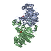 1ed8S S: Starting model for refinement C: citing same article ( |
|---|---|
| Similar structure data |
-Links
 PDBj
PDBj

- Assembly
| Deposited unit | 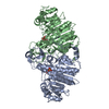
| ||||||||
|---|---|---|---|---|---|---|---|---|---|
| 1 |
| ||||||||
| Unit cell |
| ||||||||
| Details | The asymmetric unit contains biologically active dimer |
-Components
| #1: Protein | Mass: 47094.398 Da / Num. of mol.: 2 Source method: isolated from a genetically manipulated source Source: (gene. exp.) #2: Chemical | ChemComp-CO /   Mass: 58.933 Da / Num. of mol.: 6 / Source method: obtained synthetically / Formula: Co Mass: 58.933 Da / Num. of mol.: 6 / Source method: obtained synthetically / Formula: Co#3: Chemical |   Mass: 94.971 Da / Num. of mol.: 2 / Source method: obtained synthetically / Formula: PO4 Mass: 94.971 Da / Num. of mol.: 2 / Source method: obtained synthetically / Formula: PO4#4: Chemical |   Mass: 96.063 Da / Num. of mol.: 2 / Source method: obtained synthetically / Formula: SO4 Mass: 96.063 Da / Num. of mol.: 2 / Source method: obtained synthetically / Formula: SO4#5: Water | ChemComp-HOH / |  Mass: 18.015 Da / Num. of mol.: 712 / Source method: isolated from a natural source / Formula: H2O Mass: 18.015 Da / Num. of mol.: 712 / Source method: isolated from a natural source / Formula: H2OHas protein modification | Y | |
|---|
-Experimental details
-Experiment
| Experiment | Method: X-RAY DIFFRACTION / Number of used crystals: 1 |
|---|
- Sample preparation
| Crystal | Density Matthews: 3.22 Å3/Da / Density % sol: 57.2 % |
|---|---|
| Crystal grow | Temperature: 293 K / Method: vapor diffusion, hanging drop / pH: 9.5 Details: 10 mM cobalt chloride, 2.1 M Ammonium sulfate, pH 9.5, VAPOR DIFFUSION, HANGING DROP, temperature 293K |
-Data collection
| Diffraction | Mean temperature: 93 K |
|---|---|
| Diffraction source | Source: SYNCHROTRON / Site: NSLS  / Beamline: X26C / Wavelength: 1.1 Å / Beamline: X26C / Wavelength: 1.1 Å |
| Detector | Detector: AREA DETECTOR / Date: Nov 23, 2003 / Details: Monochromatic |
| Radiation | Protocol: SINGLE WAVELENGTH / Monochromatic (M) / Laue (L): M / Scattering type: x-ray |
| Radiation wavelength | Wavelength: 1.1 Å / Relative weight: 1 |
| Reflection | Resolution: 1.6→30 Å / Num. obs: 160887 / % possible obs: 99.5 % / Observed criterion σ(I): 14.9 / Redundancy: 6.9 % / Rmerge(I) obs: 0.053 |
| Reflection shell | Resolution: 1.6→1.66 Å / Redundancy: 6.5 % / Rmerge(I) obs: 0.288 / % possible all: 99.5 |
- Processing
| Software |
| ||||||||||||||||||||
|---|---|---|---|---|---|---|---|---|---|---|---|---|---|---|---|---|---|---|---|---|---|
| Refinement | Method to determine structure: MOLECULAR REPLACEMENT Starting model: pdb entry 1ED8 Resolution: 1.6→30 Å / Cross valid method: THROUGHOUT / σ(F): 0 / Stereochemistry target values: Engh & Huber
| ||||||||||||||||||||
| Refinement step | Cycle: LAST / Resolution: 1.6→30 Å
|
