 Movie
Movie Controller

[English] 日本語
 Yorodumi
- PDB-2ga3: Structure of S102T E. coli Alkaline Phosphatase-phosphate interme... -
+ Open data

- Basic information
| Entry | Database: PDB / ID: 2ga3 | ||||||
|---|---|---|---|---|---|---|---|
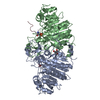
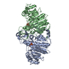
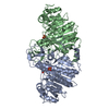
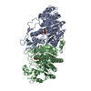
| Title | Structure of S102T E. coli Alkaline Phosphatase-phosphate intermediate at 2.20A resolution | ||||||
 Components Components | Alkaline phosphatase | ||||||
 Keywords Keywords | HYDROLASE / mutagenesis / side chain conformation / covalent intermediate / rate-determining step | ||||||
| Function / homology |  Function and homology information Function and homology informationoxidoreductase activity, acting on phosphorus or arsenic in donors / alkaline phosphatase / alkaline phosphatase activity / hydrogenase (acceptor) activity / phosphoprotein phosphatase activity / outer membrane-bounded periplasmic space / periplasmic space / magnesium ion binding / zinc ion binding Similarity search - Function | ||||||
| Biological species |  | ||||||
| Method |  X-RAY DIFFRACTION / MOLECULAR REPLACEMENT / Resolution: 2.2 Å X-RAY DIFFRACTION / MOLECULAR REPLACEMENT / Resolution: 2.2 Å | ||||||
 Authors Authors | Wang, J. / Kantrowitz, E.R. | ||||||
 Citation Citation | Journal: Protein Sci. / Year: 2006 Title: Trapping the tetrahedral intermediate in the alkaline phosphatase reaction by substitution of the active site serine with threonine. Authors: Wang, J. / Kantrowitz, E.R. | ||||||
| History |
|
- Structure visualization
| Structure viewer | Molecule: MolmilJmol/JSmol |
|---|
- Downloads & links
-Download
| PDBx/mmCIF format | 2ga3.cif.gz | 185.4 KB | Display | PDBx/mmCIF format |
|---|---|---|---|---|
| PDB format | pdb2ga3.ent.gz | 146.3 KB | Display | PDB format |
| PDBx/mmJSON format | 2ga3.json.gz | Tree view | PDBx/mmJSON format | |
| Others |  Other downloads Other downloads |
-Validation report
| Arichive directory | https://data.pdbj.org/pub/pdb/validation_reports/ga/2ga3ftp://data.pdbj.org/pub/pdb/validation_reports/ga/2ga3 | HTTPS FTP |
|---|
-Related structure data
| Related structure data |  2g9yC  1y6vS C: citing same article ( S: Starting model for refinement |
|---|---|
| Similar structure data |


-Links
 PDBj
PDBj
- Assembly
| Deposited unit | 
| ||||||||
|---|---|---|---|---|---|---|---|---|---|
| 1 |
| ||||||||
| Unit cell |
| ||||||||
| Details | The asymmetric unit contains the biologically active dimer |
-Components
| #1: Protein | Mass: 47188.402 Da / Num. of mol.: 2 / Mutation: S102(TPO) Source method: isolated from a genetically manipulated source Source: (gene. exp.) #2: Chemical | ChemComp-ZN /   Mass: 65.409 Da / Num. of mol.: 4 / Source method: obtained synthetically / Formula: Zn Mass: 65.409 Da / Num. of mol.: 4 / Source method: obtained synthetically / Formula: Zn#3: Chemical |   Mass: 24.305 Da / Num. of mol.: 2 / Source method: obtained synthetically / Formula: Mg Mass: 24.305 Da / Num. of mol.: 2 / Source method: obtained synthetically / Formula: Mg#4: Chemical |   Mass: 96.063 Da / Num. of mol.: 2 / Source method: obtained synthetically / Formula: SO4 Mass: 96.063 Da / Num. of mol.: 2 / Source method: obtained synthetically / Formula: SO4#5: Water | ChemComp-HOH / |  Mass: 18.015 Da / Num. of mol.: 495 / Source method: isolated from a natural source / Formula: H2O Mass: 18.015 Da / Num. of mol.: 495 / Source method: isolated from a natural source / Formula: H2OHas protein modification | Y | |
|---|
-Experimental details
-Experiment
| Experiment | Method: X-RAY DIFFRACTION / Number of used crystals: 1 |
|---|
- Sample preparation
| Crystal | Density Matthews: 3.21 Å3/Da / Density % sol: 61.7 % |
|---|---|
| Crystal grow | Temperature: 293 K / Method: vapor diffusion, hanging drop / pH: 9.5 Details: 2.0M ammonium sulfate, 100mM Tris, 10mM magnesium chloride, 0.01mM zinc chloride, pH 9.5, VAPOR DIFFUSION, HANGING DROP, temperature 293K |
-Data collection
| Diffraction | Mean temperature: 93 K |
|---|---|
| Diffraction source | Source: ROTATING ANODE / Type: RIGAKU RU200 / Wavelength: 1.5418 Å |
| Detector | Type: RIGAKU RAXIS IV / Detector: IMAGE PLATE / Date: Feb 26, 2004 |
| Radiation | Protocol: SINGLE WAVELENGTH / Monochromatic (M) / Laue (L): M / Scattering type: x-ray |
| Radiation wavelength | Wavelength: 1.5418 Å / Relative weight: 1 |
| Reflection | Resolution: 2.2→14.98 Å / Num. all: 61826 / Num. obs: 55150 / % possible obs: 89.2 % / Observed criterion σ(F): 0 / Observed criterion σ(I): 0 / Redundancy: 3.47 % / Rmerge(I) obs: 0.077 / Net I/σ(I): 11.3 |
| Reflection shell | Resolution: 2.2→2.28 Å / Redundancy: 3.33 % / Rmerge(I) obs: 0.352 / Num. unique all: 6110 / % possible all: 92.6 |
- Processing
| Software |
| |||||||||||||||||||||||||
|---|---|---|---|---|---|---|---|---|---|---|---|---|---|---|---|---|---|---|---|---|---|---|---|---|---|---|
| Refinement | Method to determine structure: MOLECULAR REPLACEMENT Starting model: pdb entry 1Y6V Resolution: 2.2→14.98 Å / Cross valid method: THROUGHOUT / σ(F): 0 / Stereochemistry target values: Engh & Huber
| |||||||||||||||||||||||||
| Refinement step | Cycle: LAST / Resolution: 2.2→14.98 Å
| |||||||||||||||||||||||||
| Refine LS restraints |
|
