 Movie
Movie Controller


+ Open data

- Basic information
| Entry | Database: PDB / ID: 1jwh | ||||||
|---|---|---|---|---|---|---|---|
| Title | Crystal Structure of Human Protein Kinase CK2 Holoenzyme | ||||||
 Components Components | (Casein kinase ...) x 2 | ||||||
 Keywords Keywords | TRANSFERASE / casein kinase 2 / CK2 holoenzyme / protein kinase CK2 | ||||||
| Function / homology |  Function and homology information Function and homology informationadiponectin-activated signaling pathway / positive regulation of activin receptor signaling pathway / sperm glycocalyx / endothelial tube morphogenesis / perinuclear theca / negative regulation of viral life cycle / protein kinase regulator activity / Phosphorylation and nuclear translocation of BMAL1 (ARNTL) and CLOCK / positive regulation of aggrephagy / regulation of chromosome separation ...adiponectin-activated signaling pathway / positive regulation of activin receptor signaling pathway / sperm glycocalyx / endothelial tube morphogenesis / perinuclear theca / negative regulation of viral life cycle / protein kinase regulator activity / Phosphorylation and nuclear translocation of BMAL1 (ARNTL) and CLOCK / positive regulation of aggrephagy / regulation of chromosome separation / WNT mediated activation of DVL / Condensation of Prometaphase Chromosomes / protein kinase CK2 complex / symbiont-mediated disruption of host cell PML body / Phosphorylation and nuclear translocation of the CRY:PER:kinase complex / Regulation of CDH1 posttranslational processing and trafficking to plasma membrane / Receptor Mediated Mitophagy / Synthesis of PC / Sin3-type complex / negative regulation of signal transduction by p53 class mediator / RUNX1 interacts with co-factors whose precise effect on RUNX1 targets is not known / Maturation of hRSV A proteins / negative regulation of blood vessel endothelial cell migration / positive regulation of SMAD protein signal transduction / negative regulation of apoptotic signaling pathway / negative regulation of double-strand break repair via homologous recombination / positive regulation of Wnt signaling pathway / negative regulation of proteasomal ubiquitin-dependent protein catabolic process / Signal transduction by L1 / Hsp90 protein binding / PML body / fibrillar center / Wnt signaling pathway / Regulation of PTEN stability and activity / kinase activity / positive regulation of protein catabolic process / KEAP1-NFE2L2 pathway / rhythmic process / double-strand break repair / Cooperation of PDCL (PhLP1) and TRiC/CCT in G-protein beta folding / sperm midpiece / positive regulation of cell growth / protein folding / protein-containing complex assembly / secretory granule lumen / Regulation of TP53 Activity through Phosphorylation / ficolin-1-rich granule lumen / RNA polymerase II-specific DNA-binding transcription factor binding / protein-macromolecule adaptor activity / non-specific serine/threonine protein kinase / regulation of cell cycle / negative regulation of translation / protein stabilization / negative regulation of cell population proliferation / protein domain specific binding / signaling receptor binding / protein serine kinase activity / protein serine/threonine kinase activity / chromatin binding / apoptotic process / positive regulation of cell population proliferation / Neutrophil degranulation / DNA damage response / chromatin / signal transduction / extracellular exosome / extracellular region / nucleoplasm / ATP binding / metal ion binding / identical protein binding / nucleus / plasma membrane / cytosol / cytoplasm Similarity search - Function | ||||||
| Biological species |  Homo sapiens (human) Homo sapiens (human) | ||||||
| Method |  X-RAY DIFFRACTION / SYNCHROTRON / MOLECULAR REPLACEMENT / Resolution: 3.1 Å X-RAY DIFFRACTION / SYNCHROTRON / MOLECULAR REPLACEMENT / Resolution: 3.1 Å | ||||||
 Authors Authors | Niefind, K. / Guerra, B. / Ermakowa, I. / Issinger, O.G. | ||||||
 Citation Citation | Journal: EMBO J. / Year: 2001 Title: Crystal structure of human protein kinase CK2: insights into basic properties of the CK2 holoenzyme. Authors: Niefind, K. / Guerra, B. / Ermakowa, I. / Issinger, O.G. #1: Journal: Acta Crystallogr.,Sect.D / Year: 2000Title: Crystallization and Preliminary Characterization of Crystals of Human Protein Kinase CK2 Authors: Niefind, K. / Guerra, B. / Ermakowa, I. / Issinger, O.G. | ||||||
| History |
|
- Structure visualization
| Structure viewer | Molecule: MolmilJmol/JSmol |
|---|
- Downloads & links
-Download
| PDBx/mmCIF format | 1jwh.cif.gz | 244.4 KB | Display | PDBx/mmCIF format |
|---|---|---|---|---|
| PDB format | pdb1jwh.ent.gz | 194.2 KB | Display | PDB format |
| PDBx/mmJSON format | 1jwh.json.gz | Tree view | PDBx/mmJSON format | |
| Others |  Other downloads Other downloads |
-Validation report
| Arichive directory | https://data.pdbj.org/pub/pdb/validation_reports/jw/1jwhftp://data.pdbj.org/pub/pdb/validation_reports/jw/1jwh | HTTPS FTP |
|---|
-Related structure data
| Related structure data |  1dawS S: Starting model for refinement |
|---|---|
| Similar structure data |


-Links
 PDBj
PDBj


- Assembly
| Deposited unit | 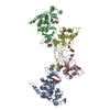
| ||||||||
|---|---|---|---|---|---|---|---|---|---|
| 1 |
| ||||||||
| Unit cell |
| ||||||||
| Details | The asymmetric unit contains one complete CK2 tetramer comprising two catalytic and two regulatory subunits. |
-Components
-Casein kinase ... , 2 types, 4 molecules ABCD
| #1: Protein | Mass: 40240.902 Da / Num. of mol.: 2 Source method: isolated from a genetically manipulated source Source: (gene. exp.) Homo sapiens (human) / Production host:  #2: Protein | Mass: 24969.412 Da / Num. of mol.: 2 Source method: isolated from a genetically manipulated source Source: (gene. exp.) Homo sapiens (human) / Production host: |
|---|
-Non-polymers , 4 types, 219 molecules 


| #3: Chemical | ChemComp-PO4 /  Mass: 94.971 Da / Num. of mol.: 7 / Source method: obtained synthetically / Formula: PO4 Mass: 94.971 Da / Num. of mol.: 7 / Source method: obtained synthetically / Formula: PO4#4: Chemical | ChemComp-ANP / |  Mass: 506.196 Da / Num. of mol.: 1 / Source method: obtained synthetically / Formula: C10H17N6O12P3 / Comment: AMP-PNP, energy-carrying molecule analogue*YM Mass: 506.196 Da / Num. of mol.: 1 / Source method: obtained synthetically / Formula: C10H17N6O12P3 / Comment: AMP-PNP, energy-carrying molecule analogue*YM#5: Chemical |  Mass: 65.409 Da / Num. of mol.: 2 / Source method: obtained synthetically / Formula: Zn Mass: 65.409 Da / Num. of mol.: 2 / Source method: obtained synthetically / Formula: Zn#6: Water | ChemComp-HOH / | Mass: 18.015 Da / Num. of mol.: 209 / Source method: isolated from a natural source / Formula: H2O |
|---|
-Experimental details
-Experiment
| Experiment | Method: X-RAY DIFFRACTION / Number of used crystals: 2 |
|---|
- Sample preparation
| Crystal | Density Matthews: 3.22 Å3/Da / Density % sol: 61.8 % Description: WHILE THE OVERALL DATA COMPLETENESS FOR THE GIVEN RESOLUTION RANGE (59.3 A - 3.1 A) WAS 98.4 % ONLY 78.7 % OF ALL POSSIBLE DATA WERE USED IN REFINEMENT WITH CNS. THE REASON FOR THIS ...Description: WHILE THE OVERALL DATA COMPLETENESS FOR THE GIVEN RESOLUTION RANGE (59.3 A - 3.1 A) WAS 98.4 % ONLY 78.7 % OF ALL POSSIBLE DATA WERE USED IN REFINEMENT WITH CNS. THE REASON FOR THIS DISCREPANCY WAS THAT THE DATA HAD NOT BEEN WILSON SCALED AFTER PROCESSING AND ALL NEGATIVE INTENSITIES HAD BEEN SET TO ZERO WHEN THE INTENSITIES HAD BEEN CONVERTED TO STRUCTURE AMPLITUDES. ALL THESE ZERO DATA WERE IGNORED DURING REFINEMENT WITH CNS. | ||||||||||||||||||||||||||||||||||||||||||||||||||||||||||||||||||||||
|---|---|---|---|---|---|---|---|---|---|---|---|---|---|---|---|---|---|---|---|---|---|---|---|---|---|---|---|---|---|---|---|---|---|---|---|---|---|---|---|---|---|---|---|---|---|---|---|---|---|---|---|---|---|---|---|---|---|---|---|---|---|---|---|---|---|---|---|---|---|---|---|
| Crystal grow | Temperature: 285 K / Method: vapor diffusion, sitting drop / pH: 9.3 Details: initial composition of the drop: 3 ul rhCK2 stock solution [5 mg/ml enzyme in 25 mM Tris/HCl, 300 mM NaCl, 1 mM dithiothreitole, pH 8.5], 1.5 ul reservoir solution [20 % (w/v) PEG3350, 200 ...Details: initial composition of the drop: 3 ul rhCK2 stock solution [5 mg/ml enzyme in 25 mM Tris/HCl, 300 mM NaCl, 1 mM dithiothreitole, pH 8.5], 1.5 ul reservoir solution [20 % (w/v) PEG3350, 200 mM dipotassium hydrogenphosphate], 3 ul 1 mM adenylyl imidodiphosphate (AMPPNP), 3 ul 2 mM magnesium chloride, 2 ul 10 % (w/v) polyethylene glycol 400 dodecylether (Thesit), pH 9.3, VAPOR DIFFUSION, SITTING DROP, temperature 285K | ||||||||||||||||||||||||||||||||||||||||||||||||||||||||||||||||||||||
| Crystal grow | *PLUS Temperature: 12 ℃ / pH: 8.5 | ||||||||||||||||||||||||||||||||||||||||||||||||||||||||||||||||||||||
| Components of the solutions | *PLUS
|
-Data collection
| Diffraction | Mean temperature: 100 K |
|---|---|
| Diffraction source | Source: SYNCHROTRON / Site: EMBL/DESY, HAMBURG  / Beamline: BW7B / Wavelength: 0.8428 Å / Beamline: BW7B / Wavelength: 0.8428 Å |
| Detector | Type: MARRESEARCH / Detector: IMAGE PLATE / Date: Jan 26, 2000 |
| Radiation | Monochromator: silicon 111 / Protocol: SINGLE WAVELENGTH / Monochromatic (M) / Laue (L): M / Scattering type: x-ray |
| Radiation wavelength | Wavelength: 0.8428 Å / Relative weight: 1 |
| Reflection | Resolution: 3.1→59.3 Å / Num. obs: 29935 / % possible obs: 98.4 % / Observed criterion σ(F): 0 / Observed criterion σ(I): 0 / Redundancy: 8.8 % / Biso Wilson estimate: 92.2 Å2 / Limit h max: 49 / Limit h min: 0 / Limit k max: 48 / Limit k min: 0 / Limit l max: 30 / Limit l min: 0 / Observed criterion F max: 2867098.76 / Observed criterion F min: 1.5 / Rsym value: 0.096 / Net I/σ(I): 16.7 |
| Reflection shell | Resolution: 3.1→3.2 Å / Redundancy: 6 % / Mean I/σ(I) obs: 2.6 / Rsym value: 42 / % possible all: 90.4 |
| Reflection | *PLUS Num. measured all: 268848 / Rmerge(I) obs: 0.096 |
| Reflection shell | *PLUS % possible obs: 90.4 % / Rmerge(I) obs: 0.42 |
- Processing
| Software |
| ||||||||||||||||||||||||||||||||||||||||||||||||||||||||||||||||||||||||||||||||||||||||||
|---|---|---|---|---|---|---|---|---|---|---|---|---|---|---|---|---|---|---|---|---|---|---|---|---|---|---|---|---|---|---|---|---|---|---|---|---|---|---|---|---|---|---|---|---|---|---|---|---|---|---|---|---|---|---|---|---|---|---|---|---|---|---|---|---|---|---|---|---|---|---|---|---|---|---|---|---|---|---|---|---|---|---|---|---|---|---|---|---|---|---|---|
| Refinement | Method to determine structure: MOLECULAR REPLACEMENT Starting model: POLY-ALANINE BACKBONE OF CK2A FROM ZEA MAYS (PDB ENTRY 1DAW) Resolution: 3.1→59.3 Å / Rfactor Rfree error: 0.011 / Occupancy max: 1 / Occupancy min: 1 / Cross valid method: THROUGHOUT / σ(F): 0 / Stereochemistry target values: MLF
| ||||||||||||||||||||||||||||||||||||||||||||||||||||||||||||||||||||||||||||||||||||||||||
| Solvent computation | Solvent model: CNS bulk solvent model used / Bsol: 27.2984 Å2 / ksol: 0.269435 e/Å3 | ||||||||||||||||||||||||||||||||||||||||||||||||||||||||||||||||||||||||||||||||||||||||||
| Displacement parameters | Biso max: 243.39 Å2 / Biso mean: 90.96 Å2 / Biso min: 3.08 Å2
| ||||||||||||||||||||||||||||||||||||||||||||||||||||||||||||||||||||||||||||||||||||||||||
| Refine Biso |
| ||||||||||||||||||||||||||||||||||||||||||||||||||||||||||||||||||||||||||||||||||||||||||
| Refine analyze |
| ||||||||||||||||||||||||||||||||||||||||||||||||||||||||||||||||||||||||||||||||||||||||||
| Refinement step | Cycle: LAST / Resolution: 3.1→59.3 Å
| ||||||||||||||||||||||||||||||||||||||||||||||||||||||||||||||||||||||||||||||||||||||||||
| Refine LS restraints |
| ||||||||||||||||||||||||||||||||||||||||||||||||||||||||||||||||||||||||||||||||||||||||||
| Refine LS restraints NCS | NCS model details: RESTRAINTS | ||||||||||||||||||||||||||||||||||||||||||||||||||||||||||||||||||||||||||||||||||||||||||
| LS refinement shell | Refine-ID: X-RAY DIFFRACTION / Total num. of bins used: 8
| ||||||||||||||||||||||||||||||||||||||||||||||||||||||||||||||||||||||||||||||||||||||||||
| Xplor file |
| ||||||||||||||||||||||||||||||||||||||||||||||||||||||||||||||||||||||||||||||||||||||||||
| Software | *PLUS Name: CNS / Version: 1 / Classification: refinement | ||||||||||||||||||||||||||||||||||||||||||||||||||||||||||||||||||||||||||||||||||||||||||
| Refinement | *PLUS Highest resolution: 3.1 Å / Lowest resolution: 60 Å / σ(F): 0 / % reflection Rfree: 4 % / Rfactor obs: 0.267 | ||||||||||||||||||||||||||||||||||||||||||||||||||||||||||||||||||||||||||||||||||||||||||
| Solvent computation | *PLUS | ||||||||||||||||||||||||||||||||||||||||||||||||||||||||||||||||||||||||||||||||||||||||||
| Displacement parameters | *PLUS Biso mean: 91 Å2 | ||||||||||||||||||||||||||||||||||||||||||||||||||||||||||||||||||||||||||||||||||||||||||
| Refine LS restraints | *PLUS
| ||||||||||||||||||||||||||||||||||||||||||||||||||||||||||||||||||||||||||||||||||||||||||
| LS refinement shell | *PLUS Rfactor Rfree: 0.458 / % reflection Rfree: 2.2 % / Rfactor Rwork: 0.458 |
