[English] 日本語
Yorodumi
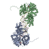
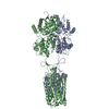
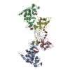
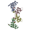
- PDB-2is4: Crystal structure of UvrD-DNA-ADPNP ternary complex -

+
Open data


ID or keywords:

Loading...

-
Basic information

Entry
Database: PDB / ID: 2is4
TitleCrystal structure of UvrD-DNA-ADPNP ternary complex
Components
  • 25-MER
  • DNA helicase II
KeywordsHYDROLASE/DNA / DNA helicase / HYDROLASE-DNA COMPLEX
Function / homology
Function and homology information


rolling circle DNA replication / single-stranded DNA-dependent ATP-dependent DNA helicase complex / mismatch repair involved in maintenance of fidelity involved in DNA-dependent DNA replication / DNA helicase complex / nucleotide-excision repair, DNA duplex unwinding / DNA translocase activity / DNA 3'-5' helicase / recombinational repair / DNA duplex unwinding / single-stranded DNA helicase activity ...rolling circle DNA replication / single-stranded DNA-dependent ATP-dependent DNA helicase complex / mismatch repair involved in maintenance of fidelity involved in DNA-dependent DNA replication / DNA helicase complex / nucleotide-excision repair, DNA duplex unwinding / DNA translocase activity / DNA 3'-5' helicase / recombinational repair / DNA duplex unwinding / single-stranded DNA helicase activity / replication fork processing / DNA unwinding involved in DNA replication / 3'-5' DNA helicase activity / SOS response / mismatch repair / DNA helicase activity / isomerase activity / nucleotide-excision repair / response to radiation / protein homodimerization activity / ATP hydrolysis activity / DNA binding / ATP binding / cytosol
Similarity search - Function
DNA helicase, ATP-dependent, UvrD type / PcrA/UvrD tudor domain / Arc Repressor Mutant, subunit A - #160 / PCRA; domain 4 / PCRA; domain 4 / DExx box DNA helicase domain superfamily / UvrD-like DNA helicase C-terminal domain profile. / UvrD-like DNA helicase, C-terminal / UvrD-like helicase C-terminal domain / UvrD/REP helicase N-terminal domain ...DNA helicase, ATP-dependent, UvrD type / PcrA/UvrD tudor domain / Arc Repressor Mutant, subunit A - #160 / PCRA; domain 4 / PCRA; domain 4 / DExx box DNA helicase domain superfamily / UvrD-like DNA helicase C-terminal domain profile. / UvrD-like DNA helicase, C-terminal / UvrD-like helicase C-terminal domain / UvrD/REP helicase N-terminal domain / UvrD-like helicase, ATP-binding domain / UvrD-like DNA helicase ATP-binding domain profile. / DNA helicase, UvrD/REP type / Arc Repressor Mutant, subunit A / P-loop containing nucleotide triphosphate hydrolases / Rossmann fold / P-loop containing nucleoside triphosphate hydrolase / Orthogonal Bundle / 3-Layer(aba) Sandwich / Mainly Alpha / Alpha Beta
Similarity search - Domain/homology
PHOSPHOAMINOPHOSPHONIC ACID-ADENYLATE ESTER / DNA / DNA (> 10) / DNA helicase II
Similarity search - Component
Biological speciesEscherichia coli (E. coli)
MethodX-RAY DIFFRACTION / SYNCHROTRON / MOLECULAR REPLACEMENT / Resolution: 2.6 Å
AuthorsYang, W. / Lee, J.Y.
CitationJournal: Cell(Cambridge,Mass.) / Year: 2006
Title: UvrD helicase unwinds DNA one base pair at a time by a two-part power stroke.
Authors: Lee, J.Y. / Yang, W.
History
DepositionOct 16, 2006Deposition site: RCSB / Processing site: RCSB
Revision 1.0Jan 9, 2007Provider: repository / Type: Initial release
Revision 1.1May 1, 2008Group: Version format compliance
Revision 1.2Jul 13, 2011Group: Version format compliance
Revision 1.3Oct 18, 2017Group: Refinement description / Category: software / Item: _software.name
Revision 1.4Oct 20, 2021Group: Database references / Derived calculations
Category: database_2 / pdbx_struct_conn_angle ...database_2 / pdbx_struct_conn_angle / struct_conn / struct_ref_seq_dif / struct_site
Item: _database_2.pdbx_DOI / _database_2.pdbx_database_accession ..._database_2.pdbx_DOI / _database_2.pdbx_database_accession / _pdbx_struct_conn_angle.ptnr1_auth_comp_id / _pdbx_struct_conn_angle.ptnr1_auth_seq_id / _pdbx_struct_conn_angle.ptnr1_label_asym_id / _pdbx_struct_conn_angle.ptnr1_label_atom_id / _pdbx_struct_conn_angle.ptnr1_label_comp_id / _pdbx_struct_conn_angle.ptnr1_label_seq_id / _pdbx_struct_conn_angle.ptnr3_auth_comp_id / _pdbx_struct_conn_angle.ptnr3_auth_seq_id / _pdbx_struct_conn_angle.ptnr3_label_asym_id / _pdbx_struct_conn_angle.ptnr3_label_atom_id / _pdbx_struct_conn_angle.ptnr3_label_comp_id / _pdbx_struct_conn_angle.ptnr3_label_seq_id / _pdbx_struct_conn_angle.value / _struct_conn.pdbx_dist_value / _struct_conn.ptnr1_auth_comp_id / _struct_conn.ptnr1_auth_seq_id / _struct_conn.ptnr1_label_asym_id / _struct_conn.ptnr1_label_atom_id / _struct_conn.ptnr1_label_comp_id / _struct_conn.ptnr1_label_seq_id / _struct_conn.ptnr2_auth_comp_id / _struct_conn.ptnr2_auth_seq_id / _struct_conn.ptnr2_label_asym_id / _struct_conn.ptnr2_label_atom_id / _struct_conn.ptnr2_label_comp_id / _struct_conn.ptnr2_label_seq_id / _struct_ref_seq_dif.details / _struct_site.pdbx_auth_asym_id / _struct_site.pdbx_auth_comp_id / _struct_site.pdbx_auth_seq_id
Revision 1.5Aug 30, 2023Group: Data collection / Refinement description
Category: chem_comp_atom / chem_comp_bond / pdbx_initial_refinement_model
Remark 300 BIOMOLECULE: 1 THIS ENTRY CONTAINS THE CRYSTALLOGRAPHIC ASYMMETRIC UNIT WHICH CONSISTS OF 4 ... BIOMOLECULE: 1 THIS ENTRY CONTAINS THE CRYSTALLOGRAPHIC ASYMMETRIC UNIT WHICH CONSISTS OF 4 CHAIN(S). THE ASSYMETRIC UNIT CONTAINS TWO BIOLOGICAL UNITS. BIOLOGICAL UNIT ONE CONTAINS CHAINS A, C (RESIDUES 1-10) AND D (RESIDUES 11-26). BIOLOGICAL UNIT TWO CONTAINS CHAINS B, C (RESIDUES 11-20) AND D (RESIDUES 22-26).

-
Structure visualization

Structure viewerMolecule:
MolmilJmol/JSmol

Downloads & links

-
Assembly

Deposited unit
C: 25-MER
D: 25-MER
A: DNA helicase II
B: DNA helicase II
hetero molecules


Theoretical massNumber of molelcules
Total (without water)172,6258
Polymers171,5644
Non-polymers1,0614
Water2,126118
1


  • Idetical with deposited unit
  • defined by author
TypeNameSymmetry operationNumber
identity operation1_555x,y,z1
Unit cell
Length a, b, c (Å)104.112, 96.779, 111.044
Angle α, β, γ (deg.)90.00, 93.85, 90.00
Int Tables number4
Space group name H-MP1211

-
Components

#1: DNA chain 25-MER


Mass: 7985.137 Da / Num. of mol.: 2 / Source method: obtained synthetically
#2: Protein DNA helicase II


Mass: 77797.078 Da / Num. of mol.: 2 / Mutation: A399V
Source method: isolated from a genetically manipulated source
Source: (gene. exp.) Escherichia coli (E. coli) / Gene: uvrD / Production host: Escherichia coli (E. coli)
References: UniProt: P03018, Hydrolases; Acting on acid anhydrides; In phosphorus-containing anhydrides
#3: Chemical ChemComp-MG / MAGNESIUM ION


Mass: 24.305 Da / Num. of mol.: 2 / Source method: obtained synthetically / Formula: Mg
#4: Chemical ChemComp-ANP / PHOSPHOAMINOPHOSPHONIC ACID-ADENYLATE ESTER


Mass: 506.196 Da / Num. of mol.: 2 / Source method: obtained synthetically / Formula: C10H17N6O12P3 / Comment: AMP-PNP, energy-carrying molecule analogue*YM
#5: Water ChemComp-HOH / water


Mass: 18.015 Da / Num. of mol.: 118 / Source method: isolated from a natural source / Formula: H2O

-
Experimental details

-
Experiment

ExperimentMethod: X-RAY DIFFRACTION / Number of used crystals: 1

-
Sample preparation

CrystalDensity Matthews: 3.24 Å3/Da / Density % sol: 62.06 %
Crystal growTemperature: 277 K / Method: vapor diffusion, sitting drop / pH: 7
Details: PEG3350, Sodium fluoride, pH 7.0, VAPOR DIFFUSION, SITTING DROP, temperature 277K
Components of the solutions
IDNameCrystal-IDSol-IDVolume3)
1PEG 335011
2Sodium Fluoride11
3H2O11
4PEG 335012
5Sodium Fluoride12Fluoride'?

-
Data collection

DiffractionMean temperature: 100 K
Diffraction sourceSource: SYNCHROTRON / Site: APS / Beamline: 22-ID / Wavelength: 1 Å
DetectorType: MAR scanner 345 mm plate / Detector: IMAGE PLATE / Date: Nov 4, 2005
RadiationMonochromator: MIRRORS / Protocol: SINGLE WAVELENGTH / Monochromatic (M) / Laue (L): M / Scattering type: x-ray
Radiation wavelengthWavelength: 1 Å / Relative weight: 1
ReflectionResolution: 2.6→50 Å / Num. all: 68018 / Num. obs: 61012 / % possible obs: 89.7 % / Observed criterion σ(F): 1 / Observed criterion σ(I): 1 / Redundancy: 3.6 % / Biso Wilson estimate: 77.04 Å2 / Rmerge(I) obs: 0.061 / Net I/σ(I): 23.3
Reflection shellResolution: 2.6→2.69 Å / Redundancy: 2.8 % / Rmerge(I) obs: 0.206 / Mean I/σ(I) obs: 4.1 / Num. unique all: 3487 / % possible all: 51.5

-
Processing

Software
NameVersionClassification
CNS1.1refinement
MAR345345DTBdata collection
HKL-2000data reduction
HKL-2000data scaling
AMoREphasing
RefinementMethod to determine structure: MOLECULAR REPLACEMENT
Starting model: PDB Entry: 2IS1
Resolution: 2.6→29.84 Å / Rfactor Rfree error: 0.003 / Data cutoff high absF: 278489.33 / Data cutoff low absF: 0 / Isotropic thermal model: RESTRAINED / Cross valid method: THROUGHOUT / σ(F): 0
RfactorNum. reflection% reflectionSelection details
Rfree0.256 5887 10.2 %RANDOM
Rwork0.215 ---
obs0.215 57871 85.3 %-
Solvent computationSolvent model: FLAT MODEL / Bsol: 42.9828 Å2 / ksol: 0.305785 e/Å3
Displacement parametersBiso mean: 79 Å2
Baniso -1Baniso -2Baniso -3
1-21.11 Å20 Å29.21 Å2
2--9.58 Å20 Å2
3----30.7 Å2
Refine analyze
FreeObs
Luzzati coordinate error0.43 Å0.37 Å
Luzzati d res low-5 Å
Luzzati sigma a0.57 Å0.52 Å
Refinement stepCycle: LAST / Resolution: 2.6→29.84 Å
ProteinNucleic acidLigandSolventTotal
Num. atoms9951 1015 64 118 11148
Refine LS restraints
Refine-IDTypeDev idealDev ideal target
X-RAY DIFFRACTIONc_bond_d0.006
X-RAY DIFFRACTIONc_bond_d_na
X-RAY DIFFRACTIONc_bond_d_prot
X-RAY DIFFRACTIONc_angle_d
X-RAY DIFFRACTIONc_angle_d_na
X-RAY DIFFRACTIONc_angle_d_prot
X-RAY DIFFRACTIONc_angle_deg1.2
X-RAY DIFFRACTIONc_angle_deg_na
X-RAY DIFFRACTIONc_angle_deg_prot
X-RAY DIFFRACTIONc_dihedral_angle_d21.2
X-RAY DIFFRACTIONc_dihedral_angle_d_na
X-RAY DIFFRACTIONc_dihedral_angle_d_prot
X-RAY DIFFRACTIONc_improper_angle_d0.82
X-RAY DIFFRACTIONc_improper_angle_d_na
X-RAY DIFFRACTIONc_improper_angle_d_prot
X-RAY DIFFRACTIONc_mcbond_it1.431.5
X-RAY DIFFRACTIONc_mcangle_it2.482
X-RAY DIFFRACTIONc_scbond_it1.932
X-RAY DIFFRACTIONc_scangle_it3.082.5
LS refinement shellResolution: 2.6→2.76 Å / Rfactor Rfree error: 0.017 / Total num. of bins used: 6
RfactorNum. reflection% reflection
Rfree0.395 520 9.6 %
Rwork0.348 4898 -
obs--48.2 %
Xplor file
Refine-IDSerial noParam fileTopol file
X-RAY DIFFRACTION1protein_rep.paramprotein.top
X-RAY DIFFRACTION2dna-rna_rep.paramdna-rna.top
X-RAY DIFFRACTION3water_rep.paramwater.top
X-RAY DIFFRACTION4ion.paramion.top
X-RAY DIFFRACTION5anp.paranp.top

+
About Yorodumi

-
News

-
Feb 9, 2022. New format data for meta-information of EMDB entries

New format data for meta-information of EMDB entries

  • Version 3 of the EMDB header file is now the official format.
  • The previous official version 1.9 will be removed from the archive.

Related info.:EMDB header

External links:wwPDB to switch to version 3 of the EMDB data model

-
Aug 12, 2020. Covid-19 info

Covid-19 info

URL: https://pdbjlvh1.pdbj.org/emnavi/covid19.php

New page: Covid-19 featured information page in EM Navigator.

Related info.:Covid-19 info / Mar 5, 2020. Novel coronavirus structure data

+
Mar 5, 2020. Novel coronavirus structure data

Novel coronavirus structure data

Related info.:Yorodumi Speices / Aug 12, 2020. Covid-19 info

External links:COVID-19 featured content - PDBj / Molecule of the Month (242):Coronavirus Proteases

+
Jan 31, 2019. EMDB accession codes are about to change! (news from PDBe EMDB page)

EMDB accession codes are about to change! (news from PDBe EMDB page)

  • The allocation of 4 digits for EMDB accession codes will soon come to an end. Whilst these codes will remain in use, new EMDB accession codes will include an additional digit and will expand incrementally as the available range of codes is exhausted. The current 4-digit format prefixed with “EMD-” (i.e. EMD-XXXX) will advance to a 5-digit format (i.e. EMD-XXXXX), and so on. It is currently estimated that the 4-digit codes will be depleted around Spring 2019, at which point the 5-digit format will come into force.
  • The EM Navigator/Yorodumi systems omit the EMD- prefix.

Related info.:Q: What is EMD? / ID/Accession-code notation in Yorodumi/EM Navigator

External links:EMDB Accession Codes are Changing Soon! / Contact to PDBj

+
Jul 12, 2017. Major update of PDB

Major update of PDB

  • wwPDB released updated PDB data conforming to the new PDBx/mmCIF dictionary.
  • This is a major update changing the version number from 4 to 5, and with Remediation, in which all the entries are updated.
  • In this update, many items about electron microscopy experimental information are reorganized (e.g. em_software).
  • Now, EM Navigator and Yorodumi are based on the updated data.

External links:wwPDB Remediation / Enriched Model Files Conforming to OneDep Data Standards Now Available in the PDB FTP Archive

-
Yorodumi

Thousand views of thousand structures

  • Yorodumi is a browser for structure data from EMDB, PDB, SASBDB, etc.
  • This page is also the successor to EM Navigator detail page, and also detail information page/front-end page for Omokage search.
  • The word "yorodu" (or yorozu) is an old Japanese word meaning "ten thousand". "mi" (miru) is to see.

Related info.:EMDB / PDB / SASBDB / Comparison of 3 databanks / Yorodumi Search / Aug 31, 2016. New EM Navigator & Yorodumi / Yorodumi Papers / Jmol/JSmol / Function and homology information / Changes in new EM Navigator and Yorodumi

Read more