 Movie
Movie Controller


+ Open data

- Basic information
| Entry | Database: PDB / ID: 1jwa | ||||||
|---|---|---|---|---|---|---|---|
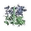
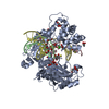
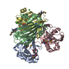
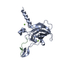
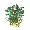
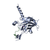
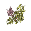
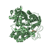
| Title | Structure of the ATP-bound MoeB-MoaD Protein Complex | ||||||
 Components Components |
| ||||||
 Keywords Keywords | LIGASE / MoeB: modified Rossmann fold / MoaD: ubiquitin-like fold | ||||||
| Function / homology |  Function and homology information Function and homology information molybdopterin-synthase adenylyltransferase / molybdopterin-synthase adenylyltransferase activity / molybdopterin synthase complex / molybdopterin adenylyltransferase complex / ubiquitin-like modifier activating enzyme activity / sulfotransferase activity / thiosulfate-cyanide sulfurtransferase activity / Mo-molybdopterin cofactor biosynthetic process / nucleotidyltransferase activity / nucleotide binding ... molybdopterin-synthase adenylyltransferase / molybdopterin-synthase adenylyltransferase activity / molybdopterin synthase complex / molybdopterin adenylyltransferase complex / ubiquitin-like modifier activating enzyme activity / sulfotransferase activity / thiosulfate-cyanide sulfurtransferase activity / Mo-molybdopterin cofactor biosynthetic process / nucleotidyltransferase activity / nucleotide binding / protein homodimerization activity / ATP binding / metal ion binding / cytosol / cytoplasm Similarity search - Function | ||||||
| Biological species |  | ||||||
| Method |  X-RAY DIFFRACTION / SYNCHROTRON / FOURIER SYNTHESIS / Resolution: 2.9 Å X-RAY DIFFRACTION / SYNCHROTRON / FOURIER SYNTHESIS / Resolution: 2.9 Å | ||||||
 Authors Authors | Lake, M.W. / Wuebbens, M.M. / Rajagopalan, K.V. / Schindelin, H. | ||||||
 Citation Citation | Journal: Nature / Year: 2001 Title: Mechanism of ubiquitin activation revealed by the structure of a bacterial MoeB-MoaD complex. Authors: Lake, M.W. / Wuebbens, M.M. / Rajagopalan, K.V. / Schindelin, H. | ||||||
| History |
|
- Structure visualization
| Structure viewer | Molecule: MolmilJmol/JSmol |
|---|
- Downloads & links
-Download
| PDBx/mmCIF format | 1jwa.cif.gz | 68.8 KB | Display | PDBx/mmCIF format |
|---|---|---|---|---|
| PDB format | pdb1jwa.ent.gz | 51.2 KB | Display | PDB format |
| PDBx/mmJSON format | 1jwa.json.gz | Tree view | PDBx/mmJSON format | |
| Others |  Other downloads Other downloads |
-Validation report
| Arichive directory | https://data.pdbj.org/pub/pdb/validation_reports/jw/1jwaftp://data.pdbj.org/pub/pdb/validation_reports/jw/1jwa | HTTPS FTP |
|---|
-Related structure data


-Links
 PDBj
PDBj


- Assembly
| Deposited unit | 
| ||||||||
|---|---|---|---|---|---|---|---|---|---|
| 1 | 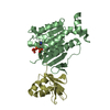
| ||||||||
| Unit cell |
| ||||||||
| Details | The asymmetric unit contains a heterodimer comprised of (1) molecule of MoeB and (1) molecule of MoaD. The heterotetramer is generated by applying: -y+1,-x+1,1/2-z |
-Components
| #1: Protein | Mass: 26741.791 Da / Num. of mol.: 1 Source method: isolated from a genetically manipulated source Source: (gene. exp.) |
|---|---|
| #2: Protein | Mass: 8764.880 Da / Num. of mol.: 1 Source method: isolated from a genetically manipulated source Source: (gene. exp.) |
| #3: Chemical | ChemComp-ATP /   Mass: 507.181 Da / Num. of mol.: 1 / Source method: obtained synthetically / Formula: C10H16N5O13P3 / Comment: ATP, energy-carrying molecule*YM Mass: 507.181 Da / Num. of mol.: 1 / Source method: obtained synthetically / Formula: C10H16N5O13P3 / Comment: ATP, energy-carrying molecule*YM |
-Experimental details
-Experiment
| Experiment | Method: X-RAY DIFFRACTION / Number of used crystals: 1 |
|---|
- Sample preparation
| Crystal | Density Matthews: 2.25 Å3/Da / Density % sol: 45.34 % | ||||||||||||||||||||||||||||||
|---|---|---|---|---|---|---|---|---|---|---|---|---|---|---|---|---|---|---|---|---|---|---|---|---|---|---|---|---|---|---|---|
| Crystal grow | Temperature: 295 K / Method: vapor diffusion, hanging drop / pH: 7.5 Details: Lithium Sulfate, HEPES, pH 7.5, VAPOR DIFFUSION, HANGING DROP, temperature 295K | ||||||||||||||||||||||||||||||
| Crystal grow | *PLUS Temperature: 4 ℃ | ||||||||||||||||||||||||||||||
| Components of the solutions | *PLUS
|
-Data collection
| Diffraction | Mean temperature: 100 K |
|---|---|
| Diffraction source | Source: SYNCHROTRON / Site: NSLS  / Beamline: X26C / Wavelength: 1.1 Å / Beamline: X26C / Wavelength: 1.1 Å |
| Detector | Type: ADSC QUANTUM 4 / Detector: CCD / Date: Apr 24, 2000 |
| Radiation | Monochromator: Si 111 CHANNEL / Protocol: SINGLE WAVELENGTH / Monochromatic (M) / Laue (L): M / Scattering type: x-ray |
| Radiation wavelength | Wavelength: 1.1 Å / Relative weight: 1 |
| Reflection | Resolution: 2.9→50 Å / Num. all: 7355 / Num. obs: 7355 / % possible obs: 98.9 % / Observed criterion σ(F): 0 / Observed criterion σ(I): 0 / Redundancy: 4.6 % / Rmerge(I) obs: 0.143 / Net I/σ(I): 9.4 |
| Reflection shell | Resolution: 2.9→3.02 Å / Rmerge(I) obs: 0.362 / Mean I/σ(I) obs: 2.4 / % possible all: 100 |
| Reflection | *PLUS |
- Processing
| Software |
| |||||||||||||||||||||||||
|---|---|---|---|---|---|---|---|---|---|---|---|---|---|---|---|---|---|---|---|---|---|---|---|---|---|---|
| Refinement | Method to determine structure: FOURIER SYNTHESIS / Resolution: 2.9→20 Å / Cross valid method: THROUGHOUT / σ(F): 0 / Stereochemistry target values: REFMAC Dictionary
| |||||||||||||||||||||||||
| Displacement parameters | Biso mean: 49.754 Å2
| |||||||||||||||||||||||||
| Refinement step | Cycle: LAST / Resolution: 2.9→20 Å
| |||||||||||||||||||||||||
| Refine LS restraints |
| |||||||||||||||||||||||||
| Software | *PLUS Name: REFMAC / Classification: refinement | |||||||||||||||||||||||||
| Refinement | *PLUS Highest resolution: 2.9 Å / Lowest resolution: 20 Å / σ(F): 0 / % reflection Rfree: 4.1 % / Rfactor obs: 0.203 | |||||||||||||||||||||||||
| Solvent computation | *PLUS | |||||||||||||||||||||||||
| Displacement parameters | *PLUS |
