 Movie
Movie Controller


+ Open data

- Basic information
| Entry | Database: PDB / ID: 5yfd | ||||||
|---|---|---|---|---|---|---|---|
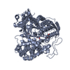
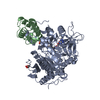
| Title | Crystal structure of a new DPP III family member | ||||||
 Components Components | Dipeptidyl peptidase 3 | ||||||
 Keywords Keywords | HYDROLASE / dipeptidyl peptidase III / aflatoxin-oxidase | ||||||
| Function / homology |  Function and homology information Function and homology informationdipeptidyl-peptidase III / metalloexopeptidase activity / dipeptidyl-peptidase activity / aminopeptidase activity / proteolysis / metal ion binding / cytoplasm Similarity search - Function | ||||||
| Biological species |  Armillaria tabescens (fungus) Armillaria tabescens (fungus) | ||||||
| Method |  X-RAY DIFFRACTION / SYNCHROTRON / Resolution: 1.76 Å X-RAY DIFFRACTION / SYNCHROTRON / Resolution: 1.76 Å | ||||||
 Authors Authors | Xu, T. / Liu, J. | ||||||
 Citation Citation | Journal: Biochem. Biophys. Res. Commun. / Year: 2017 Title: Crystal structures of Aflatoxin-oxidase from Armillariella tabescens reveal a dual activity enzyme. Authors: Xu, T. / Xie, C. / Yao, D. / Zhou, C.Z. / Liu, J. | ||||||
| History |
|
- Structure visualization
| Structure viewer | Molecule: MolmilJmol/JSmol |
|---|
- Downloads & links
-Download
| PDBx/mmCIF format | 5yfd.cif.gz | 293.6 KB | Display | PDBx/mmCIF format |
|---|---|---|---|---|
| PDB format | pdb5yfd.ent.gz | 234.3 KB | Display | PDB format |
| PDBx/mmJSON format | 5yfd.json.gz | Tree view | PDBx/mmJSON format | |
| Others |  Other downloads Other downloads |
-Validation report
| Arichive directory | https://data.pdbj.org/pub/pdb/validation_reports/yf/5yfdftp://data.pdbj.org/pub/pdb/validation_reports/yf/5yfd | HTTPS FTP |
|---|
-Related structure data


-Links
 PDBj
PDBj
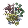
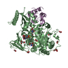
- Assembly
| Deposited unit | 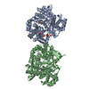
| ||||||||
|---|---|---|---|---|---|---|---|---|---|
| 1 | 
| ||||||||
| 2 | 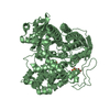
| ||||||||
| Unit cell |
|
-Components
| #1: Protein | Mass: 78181.234 Da / Num. of mol.: 2 Source method: isolated from a genetically manipulated source Source: (gene. exp.) Armillaria tabescens (fungus) / Plasmid: pET28a / Production host:  #2: Chemical | ChemComp-SO4 /   Mass: 96.063 Da / Num. of mol.: 9 / Source method: obtained synthetically / Formula: SO4 Mass: 96.063 Da / Num. of mol.: 9 / Source method: obtained synthetically / Formula: SO4#3: Chemical |   Mass: 63.546 Da / Num. of mol.: 2 / Source method: obtained synthetically / Formula: Cu Mass: 63.546 Da / Num. of mol.: 2 / Source method: obtained synthetically / Formula: Cu#4: Chemical | ChemComp-TRS / |   Mass: 122.143 Da / Num. of mol.: 1 / Source method: obtained synthetically / Formula: C4H12NO3 / Comment: pH buffer*YM Mass: 122.143 Da / Num. of mol.: 1 / Source method: obtained synthetically / Formula: C4H12NO3 / Comment: pH buffer*YM#5: Water | ChemComp-HOH / |  Mass: 18.015 Da / Num. of mol.: 806 / Source method: isolated from a natural source / Formula: H2O Mass: 18.015 Da / Num. of mol.: 806 / Source method: isolated from a natural source / Formula: H2O |
|---|
-Experimental details
-Experiment
| Experiment | Method: X-RAY DIFFRACTION / Number of used crystals: 1 |
|---|
- Sample preparation
| Crystal | Density Matthews: 2.28 Å3/Da / Density % sol: 46.15 % / Mosaicity: 0.4 ° |
|---|---|
| Crystal grow | Temperature: 293 K / Method: vapor diffusion / pH: 7.5 / Details: 0.1 M Tris-HCl pH 8.5, 2.5 M Ammonium Sulfate |
-Data collection
| Diffraction | Mean temperature: 100 K | ||||||||||||||||||||||||
|---|---|---|---|---|---|---|---|---|---|---|---|---|---|---|---|---|---|---|---|---|---|---|---|---|---|
| Diffraction source | Source: SYNCHROTRON / Site: SSRF  / Beamline: BL17U1 / Wavelength: 0.9793 Å / Beamline: BL17U1 / Wavelength: 0.9793 Å | ||||||||||||||||||||||||
| Detector | Type: MARMOSAIC 225 mm CCD / Detector: CCD / Date: May 22, 2015 | ||||||||||||||||||||||||
| Radiation | Protocol: SINGLE WAVELENGTH / Monochromatic (M) / Laue (L): M / Scattering type: x-ray | ||||||||||||||||||||||||
| Radiation wavelength | Wavelength: 0.9793 Å / Relative weight: 1 | ||||||||||||||||||||||||
| Reflection | Resolution: 1.76→69.05 Å / Num. obs: 126351 / % possible obs: 92.3 % / Redundancy: 5.5 % / CC1/2: 0.995 / Rmerge(I) obs: 0.053 / Rpim(I) all: 0.025 / Rrim(I) all: 0.059 / Net I/σ(I): 23.6 / Num. measured all: 695574 / Scaling rejects: 4566 | ||||||||||||||||||||||||
| Reflection shell | Diffraction-ID: 1
|
- Processing
| Software |
| ||||||||||||||||||||||||||||||||||||||||||||||||||||||||||||
|---|---|---|---|---|---|---|---|---|---|---|---|---|---|---|---|---|---|---|---|---|---|---|---|---|---|---|---|---|---|---|---|---|---|---|---|---|---|---|---|---|---|---|---|---|---|---|---|---|---|---|---|---|---|---|---|---|---|---|---|---|---|
| Refinement | Resolution: 1.76→58.99 Å / Cor.coef. Fo:Fc: 0.935 / Cor.coef. Fo:Fc free: 0.909 / SU B: 2.187 / SU ML: 0.072 / Cross valid method: THROUGHOUT / σ(F): 0 / ESU R: 0.133 / ESU R Free: 0.125 / Stereochemistry target values: MAXIMUM LIKELIHOOD Details: HYDROGENS HAVE BEEN ADDED IN THE RIDING POSITIONS U VALUES : REFINED INDIVIDUALLY
| ||||||||||||||||||||||||||||||||||||||||||||||||||||||||||||
| Solvent computation | Ion probe radii: 0.8 Å / Shrinkage radii: 0.8 Å / VDW probe radii: 1.2 Å / Solvent model: MASK | ||||||||||||||||||||||||||||||||||||||||||||||||||||||||||||
| Displacement parameters | Biso max: 54.62 Å2 / Biso mean: 10.284 Å2 / Biso min: 2.53 Å2
| ||||||||||||||||||||||||||||||||||||||||||||||||||||||||||||
| Refinement step | Cycle: final / Resolution: 1.76→58.99 Å
| ||||||||||||||||||||||||||||||||||||||||||||||||||||||||||||
| Refine LS restraints |
| ||||||||||||||||||||||||||||||||||||||||||||||||||||||||||||
| LS refinement shell | Resolution: 1.764→1.81 Å / Rfactor Rfree error: 0 / Total num. of bins used: 20
|
