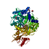
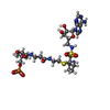
Entry Database : PDB / ID : 6o6eTitle Crystal structure of PltF trapped with PltL using a proline adenosine vinylsulfonamide inhibitor L-proline--[L-prolyl-carrier protein] ligase Peptidyl carrier protein PltL Keywords / / / / / / Function / homology Function Domain/homology Component
/ / / / / / / / / / / / / / / / / / / / / / / / / / / / / / / / / / / / / / / / / Biological species Pseudomonas protegens Pf-5 (bacteria)Method / / / Resolution : 2.14 Å Authors Corpuz, J.C. / Podust, L.M. Funding support Organization Grant number Country National Institutes of Health/National Human Genome Research Institute (NIH/NHGRI) T32 GM008326 National Institutes of Health/National Human Genome Research Institute (NIH/NHGRI) R01 GM095970
Journal : Rsc Chem Biol / Year : 2020Title : Dynamic visualization of type II peptidyl carrier protein recognition in pyoluteorin biosynthesis.Authors : Corpuz, J.C. / Podust, L.M. / Davis, T.D. / Jaremko, M.J. / Burkart, M.D. History Deposition Mar 6, 2019 Deposition site / Processing site Revision 1.0 Apr 8, 2020 Provider / Type Revision 1.1 Jun 17, 2020 Group / Category / citation_authorItem / _citation.title / _citation.yearRevision 2.0 Jul 22, 2020 Group Advisory / Atomic model ... Advisory / Atomic model / Author supporting evidence / Data collection / Database references / Derived calculations / Polymer sequence / Refinement description / Source and taxonomy / Structure summary Category atom_site / citation ... atom_site / citation / computing / entity / entity_name_com / entity_poly / entity_poly_seq / entity_src_gen / pdbx_entity_instance_feature / pdbx_entry_details / pdbx_nonpoly_scheme / pdbx_poly_seq_scheme / pdbx_struct_assembly_prop / pdbx_unobs_or_zero_occ_atoms / pdbx_unobs_or_zero_occ_residues / refine / refine_hist / refine_ls_restr / refine_ls_shell / reflns / reflns_shell / struct_keywords / struct_ref / struct_ref_seq / struct_ref_seq_dif Item _citation.pdbx_database_id_DOI / _citation.year ... _citation.pdbx_database_id_DOI / _citation.year / _entity.formula_weight / _entity.pdbx_description / _entity.pdbx_ec / _entity_poly.pdbx_seq_one_letter_code / _entity_poly.pdbx_seq_one_letter_code_can / _entity_src_gen.pdbx_end_seq_num / _pdbx_nonpoly_scheme.auth_mon_id / _pdbx_nonpoly_scheme.auth_seq_num / _pdbx_struct_assembly_prop.value / _refine.B_iso_max / _refine.B_iso_mean / _refine.B_iso_min / _refine.ls_R_factor_R_free / _refine.ls_R_factor_R_work / _refine.ls_R_factor_obs / _refine.ls_percent_reflns_obs / _refine_hist.cycle_id / _refine_hist.number_atoms_total / _refine_hist.pdbx_B_iso_mean_ligand / _refine_hist.pdbx_B_iso_mean_solvent / _refine_hist.pdbx_number_atoms_protein / _refine_hist.pdbx_number_residues_total / _refine_ls_shell.R_factor_R_free_error / _refine_ls_shell.d_res_high / _refine_ls_shell.d_res_low / _refine_ls_shell.number_reflns_all / _reflns.observed_criterion_sigma_F / _reflns.pdbx_CC_half / _reflns.pdbx_CC_star / _reflns.pdbx_Rrim_I_all / _reflns_shell.number_unique_obs / _reflns_shell.pdbx_CC_half / _reflns_shell.pdbx_CC_star / _reflns_shell.pdbx_Rrim_I_all / _reflns_shell.pdbx_redundancy / _struct_keywords.pdbx_keywords / _struct_keywords.text / _struct_ref.db_code / _struct_ref.pdbx_seq_one_letter_code / _struct_ref_seq.db_align_end / _struct_ref_seq.pdbx_auth_seq_align_end / _struct_ref_seq.seq_align_end Description / Details / Provider / Type Revision 2.1 Nov 11, 2020 Group / Category Item _citation.journal_abbrev / _citation.journal_id_CSD ... _citation.journal_abbrev / _citation.journal_id_CSD / _citation.pdbx_database_id_DOI / _citation.year Revision 2.2 Dec 23, 2020 Group / Category / citation_authorItem _citation.country / _citation.journal_abbrev ... _citation.country / _citation.journal_abbrev / _citation.journal_id_ISSN / _citation.journal_volume / _citation.page_first / _citation.page_last / _citation.pdbx_database_id_DOI / _citation.pdbx_database_id_PubMed / _citation.title / _citation_author.identifier_ORCID Revision 3.0 Jul 20, 2022 Group Advisory / Atomic model ... Advisory / Atomic model / Author supporting evidence / Data collection / Database references / Derived calculations / Non-polymer description / Refinement description / Source and taxonomy / Structure summary Category atom_site / chem_comp ... atom_site / chem_comp / database_2 / entity / entity_src_gen / pdbx_contact_author / pdbx_entity_instance_feature / pdbx_entity_nonpoly / pdbx_entry_details / pdbx_nonpoly_scheme / pdbx_struct_assembly_prop / pdbx_validate_close_contact / pdbx_validate_torsion / refine / refine_hist / refine_ls_restr / refine_ls_shell / reflns / software / struct_conf / struct_conn / struct_sheet_range / struct_site / struct_site_gen Item _atom_site.B_iso_or_equiv / _atom_site.Cartn_x ... _atom_site.B_iso_or_equiv / _atom_site.Cartn_x / _atom_site.Cartn_y / _atom_site.Cartn_z / _atom_site.auth_comp_id / _atom_site.auth_seq_id / _atom_site.label_comp_id / _chem_comp.formula / _chem_comp.formula_weight / _chem_comp.id / _chem_comp.mon_nstd_flag / _chem_comp.name / _chem_comp.type / _database_2.pdbx_DOI / _database_2.pdbx_database_accession / _entity.pdbx_description / _entity_src_gen.pdbx_gene_src_scientific_name / _pdbx_entity_nonpoly.comp_id / _pdbx_entity_nonpoly.name / _pdbx_entry_details.has_ligand_of_interest / _pdbx_nonpoly_scheme.auth_mon_id / _pdbx_nonpoly_scheme.auth_seq_num / _pdbx_nonpoly_scheme.mon_id / _pdbx_nonpoly_scheme.pdb_mon_id / _pdbx_nonpoly_scheme.pdb_seq_num / _pdbx_struct_assembly_prop.value / _refine.B_iso_max / _refine.B_iso_mean / _refine.B_iso_min / _refine.aniso_B[1][1] / _refine.aniso_B[1][2] / _refine.aniso_B[2][2] / _refine.aniso_B[3][3] / _refine.correlation_coeff_Fo_to_Fc / _refine.correlation_coeff_Fo_to_Fc_free / _refine.details / _refine.ls_R_factor_R_free / _refine.ls_R_factor_R_work / _refine.ls_R_factor_obs / _refine.ls_d_res_low / _refine.ls_number_reflns_obs / _refine.ls_percent_reflns_obs / _refine.overall_SU_B / _refine.overall_SU_ML / _refine.pdbx_ls_sigma_F / _refine.pdbx_overall_ESU_R / _refine.pdbx_overall_ESU_R_Free / _refine.pdbx_stereochemistry_target_values / _refine.solvent_model_details / _refine_hist.d_res_low / _refine_hist.pdbx_B_iso_mean_ligand / _refine_hist.pdbx_B_iso_mean_solvent / _refine_ls_shell.R_factor_R_free / _refine_ls_shell.R_factor_R_work / _refine_ls_shell.d_res_high / _refine_ls_shell.number_reflns_R_work / _refine_ls_shell.number_reflns_all / _refine_ls_shell.percent_reflns_obs / _reflns.d_resolution_low / _software.version / _struct_conn.pdbx_dist_value / _struct_conn.ptnr2_auth_comp_id / _struct_conn.ptnr2_auth_seq_id / _struct_conn.ptnr2_label_comp_id / _struct_sheet_range.end_auth_comp_id / _struct_sheet_range.end_auth_seq_id / _struct_sheet_range.end_label_comp_id / _struct_sheet_range.end_label_seq_id Description Details Provider / Type Revision 3.1 Oct 11, 2023 Group / Refinement descriptionCategory / chem_comp_bond / pdbx_initial_refinement_modelRevision 3.2 Oct 23, 2024 Group / Category / pdbx_modification_feature / Item
Show all Show less
 Movie
Movie Controller

 Yorodumi
 Open data
 Basic information
 Components
 Keywords
 Function and homology information
 Pseudomonas protegens Pf-5 (bacteria)
 X-RAY DIFFRACTION /
  Authors
 United States, 2items
  Citation
 Structure visualization
 Downloads & links
 Other downloads
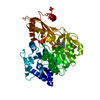


 PDBj
PDBj


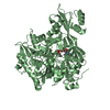
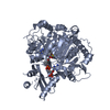
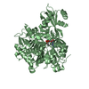
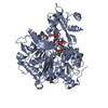
 Assembly
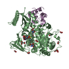


 Mass: 46.025 Da / Num. of mol.: 13 / Source method: obtained synthetically / Formula: CH2O2
 Mass: 122.143 Da / Num. of mol.: 1 / Source method: obtained synthetically / Formula: C4H12NO3 / Comment: pH buffer*YM
 Mass: 104.061 Da / Num. of mol.: 2 / Source method: obtained synthetically / Formula: C3H4O4
 Mass: 765.796 Da / Num. of mol.: 1 / Source method: obtained synthetically / Formula: C27H44N9O11PS2 / Feature type: SUBJECT OF INVESTIGATION
 Sample preparation
 Processing