 Movie
Movie Controller

[English] 日本語
 Yorodumi
- PDB-5gxd: Structure of acryloyl-CoA lyase PrpE from Dinoroseobacter shibae ... -
+ Open data

- Basic information
| Entry | Database: PDB / ID: 5gxd | ||||||
|---|---|---|---|---|---|---|---|
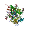
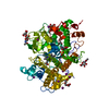
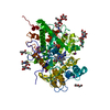
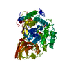
| Title | Structure of acryloyl-CoA lyase PrpE from Dinoroseobacter shibae DFL 12 | ||||||
 Components Components | AMP-dependent synthetase and ligase | ||||||
 Keywords Keywords | LYASE / acryloyl-CoA / acrylate | ||||||
| Function / homology |  Function and homology information Function and homology informationpropionate-CoA ligase activity / Ligases; Forming carbon-sulfur bonds; Acid-thiol ligases / nucleotide binding Similarity search - Function | ||||||
| Biological species |  Dinoroseobacter shibae (bacteria) Dinoroseobacter shibae (bacteria) | ||||||
| Method |  X-RAY DIFFRACTION / SYNCHROTRON / MOLECULAR REPLACEMENT / Resolution: 2.001 Å X-RAY DIFFRACTION / SYNCHROTRON / MOLECULAR REPLACEMENT / Resolution: 2.001 Å | ||||||
 Authors Authors | Zhang, Y.Z. / Wang, P. / Cao, H.Y. | ||||||
| Funding support |  China, 1items China, 1items
| ||||||
 Citation Citation | Journal: To Be Published Title: Structure of acryloyl-CoA lyase PrpE from Dinoroseobacter shibae DFL 12 Authors: Zhang, Y.Z. / Wang, P. / Cao, H.Y. | ||||||
| History |
|
- Structure visualization
| Structure viewer | Molecule: MolmilJmol/JSmol |
|---|
- Downloads & links
-Download
| PDBx/mmCIF format | 5gxd.cif.gz | 149.8 KB | Display | PDBx/mmCIF format |
|---|---|---|---|---|
| PDB format | pdb5gxd.ent.gz | 113.5 KB | Display | PDB format |
| PDBx/mmJSON format | 5gxd.json.gz | Tree view | PDBx/mmJSON format | |
| Others |  Other downloads Other downloads |
-Validation report
| Arichive directory | https://data.pdbj.org/pub/pdb/validation_reports/gx/5gxdftp://data.pdbj.org/pub/pdb/validation_reports/gx/5gxd | HTTPS FTP |
|---|
-Related structure data
| Related structure data |  2p2bS S: Starting model for refinement |
|---|---|
| Similar structure data |


-Links
 PDBj
PDBj


- Assembly
| Deposited unit | 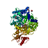
| ||||||||
|---|---|---|---|---|---|---|---|---|---|
| 1 |
| ||||||||
| Unit cell |
|
-Components
-Protein , 1 types, 1 molecules A
| #1: Protein | Mass: 68485.727 Da / Num. of mol.: 1 Source method: isolated from a genetically manipulated source Source: (gene. exp.) Dinoroseobacter shibae (strain DSM 16493 / NCIMB 14021 / DFL 12) (bacteria)Strain: DSM 16493 / NCIMB 14021 / DFL 12 / Gene: Dshi_0825 / Production host: References: UniProt: A8LRC0, Ligases; Forming carbon-sulfur bonds; Acid-thiol ligases |
|---|
-Non-polymers , 5 types, 544 molecules 


| #2: Chemical | ChemComp-AMP /  Mass: 347.221 Da / Num. of mol.: 1 / Source method: obtained synthetically / Formula: C10H14N5O7P / Comment: AMP*YM Mass: 347.221 Da / Num. of mol.: 1 / Source method: obtained synthetically / Formula: C10H14N5O7P / Comment: AMP*YM | ||
|---|---|---|---|
| #3: Chemical | ChemComp-AKR /  Mass: 72.063 Da / Num. of mol.: 1 / Source method: obtained synthetically / Formula: C3H4O2 Mass: 72.063 Da / Num. of mol.: 1 / Source method: obtained synthetically / Formula: C3H4O2 | ||
| #4: Chemical | ChemComp-COA /  Mass: 767.534 Da / Num. of mol.: 1 / Source method: obtained synthetically / Formula: C21H36N7O16P3S Mass: 767.534 Da / Num. of mol.: 1 / Source method: obtained synthetically / Formula: C21H36N7O16P3S | ||
| #5: Chemical | ChemComp-GOL /  Mass: 92.094 Da / Num. of mol.: 5 / Source method: obtained synthetically / Formula: C3H8O3 Mass: 92.094 Da / Num. of mol.: 5 / Source method: obtained synthetically / Formula: C3H8O3#6: Water | ChemComp-HOH / | Mass: 18.015 Da / Num. of mol.: 536 / Source method: isolated from a natural source / Formula: H2O |
-Experimental details
-Experiment
| Experiment | Method: X-RAY DIFFRACTION / Number of used crystals: 1 |
|---|
- Sample preparation
| Crystal | Density Matthews: 2.79 Å3/Da / Density % sol: 55.9 % |
|---|---|
| Crystal grow | Temperature: 293 K / Method: vapor diffusion, hanging drop Details: 1.8M Ammonium sulfate, 0.1M BIS-TRIS pH 6.5, 2% polyethylene glycol(PEG) monomethyl ether 550 |
-Data collection
| Diffraction | Mean temperature: 100 K |
|---|---|
| Diffraction source | Source: SYNCHROTRON / Site: SSRF / Beamline: BL17U / Wavelength: 0.9791 Å |
| Detector | Type: ADSC QUANTUM 315r / Detector: CCD / Date: Oct 12, 2015 |
| Radiation | Protocol: SINGLE WAVELENGTH / Monochromatic (M) / Laue (L): M / Scattering type: x-ray |
| Radiation wavelength | Wavelength: 0.9791 Å / Relative weight: 1 |
| Reflection | Resolution: 2.0008→50 Å / Num. obs: 50809 / % possible obs: 99.6 % / Redundancy: 3.7 % / Rmerge(I) obs: 0.071 / Net I/σ(I): 28.07 |
| Reflection shell | Resolution: 2.001→2.073 Å / Redundancy: 3.7 % / Rmerge(I) obs: 0.287 / Num. unique obs: 4985 / % possible all: 99 |
- Processing
| Software |
| |||||||||||||||||||||||||||||||||||||||||||||||||||||||||||||||||||||||||||||||||||||||||||||||||||||||||||||||||||||||||||||||||||||
|---|---|---|---|---|---|---|---|---|---|---|---|---|---|---|---|---|---|---|---|---|---|---|---|---|---|---|---|---|---|---|---|---|---|---|---|---|---|---|---|---|---|---|---|---|---|---|---|---|---|---|---|---|---|---|---|---|---|---|---|---|---|---|---|---|---|---|---|---|---|---|---|---|---|---|---|---|---|---|---|---|---|---|---|---|---|---|---|---|---|---|---|---|---|---|---|---|---|---|---|---|---|---|---|---|---|---|---|---|---|---|---|---|---|---|---|---|---|---|---|---|---|---|---|---|---|---|---|---|---|---|---|---|---|---|
| Refinement | Method to determine structure: MOLECULAR REPLACEMENT Starting model: 2P2B Resolution: 2.001→31.316 Å / SU ML: 0.17 / Cross valid method: FREE R-VALUE / σ(F): 1.34 / Phase error: 20.79 / Stereochemistry target values: ML
| |||||||||||||||||||||||||||||||||||||||||||||||||||||||||||||||||||||||||||||||||||||||||||||||||||||||||||||||||||||||||||||||||||||
| Solvent computation | Shrinkage radii: 0.9 Å / VDW probe radii: 1.11 Å / Solvent model: FLAT BULK SOLVENT MODEL | |||||||||||||||||||||||||||||||||||||||||||||||||||||||||||||||||||||||||||||||||||||||||||||||||||||||||||||||||||||||||||||||||||||
| Refinement step | Cycle: LAST / Resolution: 2.001→31.316 Å
| |||||||||||||||||||||||||||||||||||||||||||||||||||||||||||||||||||||||||||||||||||||||||||||||||||||||||||||||||||||||||||||||||||||
| Refine LS restraints |
| |||||||||||||||||||||||||||||||||||||||||||||||||||||||||||||||||||||||||||||||||||||||||||||||||||||||||||||||||||||||||||||||||||||
| LS refinement shell |
|
