 Movie
Movie Controller

[English] 日本語
 Yorodumi
- PDB-1lla: CRYSTAL STRUCTURE OF DEOXYGENATED LIMULUS POLYPHEMUS SUBUNIT II H... -
+ Open data

- Basic information
| Entry | Database: PDB / ID: 1lla | ||||||
|---|---|---|---|---|---|---|---|
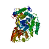
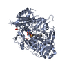
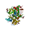
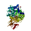
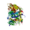
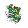
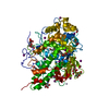
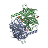
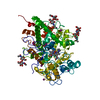
| Title | CRYSTAL STRUCTURE OF DEOXYGENATED LIMULUS POLYPHEMUS SUBUNIT II HEMOCYANIN AT 2.18 ANGSTROMS RESOLUTION: CLUES FOR A MECHANISM FOR ALLOSTERIC REGULATION | ||||||
 Components Components | HEMOCYANIN (SUBUNIT TYPE II) | ||||||
 Keywords Keywords | OXYGEN TRANSPORT | ||||||
| Function / homology |  Function and homology information Function and homology informationchloride ion binding / oxygen transport / oxygen carrier activity / oxidoreductase activity / copper ion binding / extracellular region Similarity search - Function | ||||||
| Biological species |  Limulus polyphemus (Atlantic horseshoe crab) Limulus polyphemus (Atlantic horseshoe crab) | ||||||
| Method |  X-RAY DIFFRACTION / Resolution: 2.18 Å X-RAY DIFFRACTION / Resolution: 2.18 Å | ||||||
 Authors Authors | Hazes, B. / Hol, W.G.J. | ||||||
 Citation Citation | Journal: Protein Sci. / Year: 1993 Title: Crystal structure of deoxygenated Limulus polyphemus subunit II hemocyanin at 2.18 A resolution: clues for a mechanism for allosteric regulation. Authors: Hazes, B. / Magnus, K.A. / Bonaventura, C. / Bonaventura, J. / Dauter, Z. / Kalk, K.H. / Hol, W.G. #1: Journal: Proteins / Year: 1991Title: Hexamers of Subunit II from Limulus Hemocyanin (A 48-mer) Have the Same Quaternary Structure as Whole Panulirus Hemocyanin Molecules Authors: Magnus, K.A. / Lattman, E.E. / Volbeda, A. / Hol, W.G.J. #2: Journal: J.Mol.Biol. / Year: 1989Title: Crystal Structure of Hexameric Haemocyanin from Panulirus Interruptus Refined at 3.2 Angstroms Resolution Authors: Volbeda, A. / Hol, W.G.J. #3: Journal: J.Biol.Chem. / Year: 1986Title: Structure of Hemocyanin II from the Horshoe Crab Limulus Polyphemus Authors: Nakashima, H. / Behrens, P.Q. / Moore, M.D. / Yokota, E. / Riggs, A.F. | ||||||
| History |
|
- Structure visualization
| Structure viewer | Molecule: MolmilJmol/JSmol |
|---|
- Downloads & links
-Download
| PDBx/mmCIF format | 1lla.cif.gz | 141.4 KB | Display | PDBx/mmCIF format |
|---|---|---|---|---|
| PDB format | pdb1lla.ent.gz | 109.5 KB | Display | PDB format |
| PDBx/mmJSON format | 1lla.json.gz | Tree view | PDBx/mmJSON format | |
| Others |  Other downloads Other downloads |
-Validation report
| Arichive directory | https://data.pdbj.org/pub/pdb/validation_reports/ll/1llaftp://data.pdbj.org/pub/pdb/validation_reports/ll/1lla | HTTPS FTP |
|---|
-Related structure data


-Links
 PDBj
PDBj- Assembly
| Deposited unit | 
| ||||||||
|---|---|---|---|---|---|---|---|---|---|
| 1 |
| ||||||||
| Unit cell |
| ||||||||
| Atom site foot note | 1: RESIDUE PRO 599 IS A CIS PROLINE. |
-Components
| #1: Protein | Mass: 72757.695 Da / Num. of mol.: 1 Source method: isolated from a genetically manipulated source Source: (gene. exp.) Limulus polyphemus (Atlantic horseshoe crab)References: UniProt: P04253 | ||||||||||||
|---|---|---|---|---|---|---|---|---|---|---|---|---|---|
| #2: Chemical |   Mass: 63.546 Da / Num. of mol.: 2 / Source method: obtained synthetically / Formula: Cu Mass: 63.546 Da / Num. of mol.: 2 / Source method: obtained synthetically / Formula: Cu#3: Chemical | ChemComp-NA / |   Mass: 22.990 Da / Num. of mol.: 1 / Source method: obtained synthetically / Formula: Na Mass: 22.990 Da / Num. of mol.: 1 / Source method: obtained synthetically / Formula: Na#4: Chemical | ChemComp-CL / |   Mass: 35.453 Da / Num. of mol.: 1 / Source method: obtained synthetically / Formula: Cl Mass: 35.453 Da / Num. of mol.: 1 / Source method: obtained synthetically / Formula: Cl#5: Water | ChemComp-HOH / |  Mass: 18.015 Da / Num. of mol.: 332 / Source method: isolated from a natural source / Formula: H2O Mass: 18.015 Da / Num. of mol.: 332 / Source method: isolated from a natural source / Formula: H2OCompound details | BASED ON OXYGEN BINDING STUDIES THAT MIMIC THE CRYSTALLIZATION CONDITIONS THE STRUCTURE IS BELIEVED ...BASED ON OXYGEN BINDING STUDIES THAT MIMIC THE CRYSTALLIZ | Has protein modification | Y | Sequence details | SEQUENCE ADVISORY NOTICE: THE SEQUENCE HAS BEEN CHANGED IN THREE POSITIONS COMPARED TO THE ...SEQUENCE ADVISORY NOTICE: THE SEQUENCE HAS BEEN CHANGED IN THREE POSITIONS COMPARED TO THE PUBLISHED SEQUENCE (NAKASHIMA, 1986). DIFFERENCE | |
-Experimental details
-Experiment
| Experiment | Method: X-RAY DIFFRACTION |
|---|
- Sample preparation
| Crystal | Density Matthews: 2.59 Å3/Da / Density % sol: 52.59 % | ||||||||||||||||||||||||||||||||||||||||||
|---|---|---|---|---|---|---|---|---|---|---|---|---|---|---|---|---|---|---|---|---|---|---|---|---|---|---|---|---|---|---|---|---|---|---|---|---|---|---|---|---|---|---|---|
| Crystal grow | *PLUS pH: 6 / Method: vapor diffusion, hanging dropDetails: taken from mangus, K.A. et al (1991). Proteins Struct. Funct. Genet., 9, 240-247. | ||||||||||||||||||||||||||||||||||||||||||
| Components of the solutions | *PLUS
|
-Data collection
| Radiation | Scattering type: x-ray |
|---|---|
| Radiation wavelength | Relative weight: 1 |
| Reflection | *PLUS Highest resolution: 2.18 Å / % possible obs: 97 % / Num. measured all: 38255 / Rmerge(I) obs: 0.046 |
- Processing
| Software |
| ||||||||||||||||||||||||||||||||||||||||||||||||||||||||||||
|---|---|---|---|---|---|---|---|---|---|---|---|---|---|---|---|---|---|---|---|---|---|---|---|---|---|---|---|---|---|---|---|---|---|---|---|---|---|---|---|---|---|---|---|---|---|---|---|---|---|---|---|---|---|---|---|---|---|---|---|---|---|
| Refinement | Resolution: 2.18→10 Å / Rfactor Rwork: 0.174 / Rfactor obs: 0.174 / σ(F): 0 | ||||||||||||||||||||||||||||||||||||||||||||||||||||||||||||
| Refinement step | Cycle: LAST / Resolution: 2.18→10 Å
| ||||||||||||||||||||||||||||||||||||||||||||||||||||||||||||
| Refine LS restraints |
| ||||||||||||||||||||||||||||||||||||||||||||||||||||||||||||
| Software | *PLUS Name: X-PLOR / Classification: refinement | ||||||||||||||||||||||||||||||||||||||||||||||||||||||||||||
| Refinement | *PLUS Highest resolution: 2.18 Å / Lowest resolution: 10 Å / σ(F): 0 / Rfactor all: 0.174 | ||||||||||||||||||||||||||||||||||||||||||||||||||||||||||||
| Solvent computation | *PLUS | ||||||||||||||||||||||||||||||||||||||||||||||||||||||||||||
| Displacement parameters | *PLUS | ||||||||||||||||||||||||||||||||||||||||||||||||||||||||||||
| Refine LS restraints | *PLUS Type: x_angle_d / Dev ideal: 2.9 |
