- PDB-1ll1: HYDROXO BRIDGE MET FORM HEMOCYANIN FROM LIMULUS -
+
Open data
ID or keywords:
Loading...
-
Basic information
Entry
Database: PDB / ID: 1ll1
Title
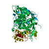
HYDROXO BRIDGE MET FORM HEMOCYANIN FROM LIMULUS
Components
METCYANIN II
Keywords
OXYGEN TRANSPORT / RESPIRATORY PROTEIN / GLYCOPROTEIN
Function / homology
Function and homology information
chloride ion binding / oxygen transport / oxygen carrier activity / oxidoreductase activity / copper ion binding / extracellular region Similarity search - Function
Resolution: 2.55→60 Å / Rfactor Rfree error: 0.02 / Data cutoff high absF: 10000000 / Data cutoff low absF: 0.1 / Isotropic thermal model: RESTRAINED / Cross valid method: THROUGHOUT / σ(F): 2 / Details: THERE ARE SEVERAL RESIDUE
Rfactor
Num. reflection
% reflection
Selection details
Rfree
0.23
2276
10 %
RANDOM
Rwork
0.167
-
-
-
obs
0.167
21960
88.6 %
-
Displacement parameters
Biso mean: 29.8 Å2
Baniso -1
Baniso -2
Baniso -3
1-
17.434 Å2
7.064 Å2
0 Å2
2-
-
17.434 Å2
0 Å2
3-
-
-
17.24 Å2
Refine analyze
Free
Obs
Luzzati coordinate error
0.33 Å
0.25 Å
Luzzati d res low
-
5 Å
Luzzati sigma a
0.35 Å
0.28 Å
Refinement step
Cycle: LAST / Resolution: 2.55→60 Å
Protein
Nucleic acid
Ligand
Solvent
Total
Num. atoms
4763
0
4
135
4902
Refine LS restraints
Refine-ID
Type
Dev ideal
Dev ideal target
X-RAY DIFFRACTION
x_bond_d
0.006
X-RAY DIFFRACTION
x_bond_d_na
X-RAY DIFFRACTION
x_bond_d_prot
X-RAY DIFFRACTION
x_angle_d
X-RAY DIFFRACTION
x_angle_d_na
X-RAY DIFFRACTION
x_angle_d_prot
X-RAY DIFFRACTION
x_angle_deg
1.6
X-RAY DIFFRACTION
x_angle_deg_na
X-RAY DIFFRACTION
x_angle_deg_prot
X-RAY DIFFRACTION
x_dihedral_angle_d
23.1
X-RAY DIFFRACTION
x_dihedral_angle_d_na
X-RAY DIFFRACTION
x_dihedral_angle_d_prot
X-RAY DIFFRACTION
x_improper_angle_d
1
X-RAY DIFFRACTION
x_improper_angle_d_na
X-RAY DIFFRACTION
x_improper_angle_d_prot
X-RAY DIFFRACTION
x_mcbond_it
2.76
1.5
X-RAY DIFFRACTION
x_mcangle_it
4.242
2
X-RAY DIFFRACTION
x_scbond_it
4.297
2
X-RAY DIFFRACTION
x_scangle_it
6.195
2.5
LS refinement shell
Resolution: 2.55→2.67 Å / Rfactor Rfree error: 0.04 / Total num. of bins used: 8
Rfactor
Num. reflection
% reflection
Rfree
0.279
156
11.1 %
Rwork
0.248
1241
-
obs
-
-
45.6 %
Xplor file
Refine-ID
Serial no
Param file
Topol file
X-RAY DIFFRACTION
1
PARHCSDX.PRO
TOPHCSDX.PRO
X-RAY DIFFRACTION
2
+
About Yorodumi
-
News
-
Feb 9, 2022. New format data for meta-information of EMDB entries
New format data for meta-information of EMDB entries
Version 3 of the EMDB header file is now the official format.
The previous official version 1.9 will be removed from the archive.
In the structure databanks used in Yorodumi, some data are registered as the other names, "COVID-19 virus" and "2019-nCoV". Here are the details of the virus and the list of structure data.
Jan 31, 2019. EMDB accession codes are about to change! (news from PDBe EMDB page)
EMDB accession codes are about to change! (news from PDBe EMDB page)
The allocation of 4 digits for EMDB accession codes will soon come to an end. Whilst these codes will remain in use, new EMDB accession codes will include an additional digit and will expand incrementally as the available range of codes is exhausted. The current 4-digit format prefixed with “EMD-” (i.e. EMD-XXXX) will advance to a 5-digit format (i.e. EMD-XXXXX), and so on. It is currently estimated that the 4-digit codes will be depleted around Spring 2019, at which point the 5-digit format will come into force.
The EM Navigator/Yorodumi systems omit the EMD- prefix.
Related info.:Q: What is EMD? / ID/Accession-code notation in Yorodumi/EM Navigator
Yorodumi is a browser for structure data from EMDB, PDB, SASBDB, etc.
This page is also the successor to EM Navigator detail page, and also detail information page/front-end page for Omokage search.
The word "yorodu" (or yorozu) is an old Japanese word meaning "ten thousand". "mi" (miru) is to see.
Related info.:EMDB / PDB / SASBDB / Comparison of 3 databanks / Yorodumi Search / Aug 31, 2016. New EM Navigator & Yorodumi / Yorodumi Papers / Jmol/JSmol / Function and homology information / Changes in new EM Navigator and Yorodumi
 Movie
Movie Controller


 Open data
 Basic information
 Components
 Keywords
 Function and homology information
 Limulus polyphemus (Atlantic horseshoe crab)
 X-RAY DIFFRACTION /
  Authors
 Citation
 Structure visualization
 Downloads & links
 Other downloads


 PDBj
PDBj Assembly

 Mass: 63.546 Da / Num. of mol.: 2 / Source method: obtained synthetically / Formula: Cu
 Mass: 35.453 Da / Num. of mol.: 1 / Source method: obtained synthetically / Formula: Cl
 Mass: 18.015 Da / Num. of mol.: 137 / Source method: isolated from a natural source / Formula: H2O
 Sample preparation
 Processing