 Movie
Movie Controller


+ Open data

- Basic information
| Entry | Database: PDB / ID: 5egy | |||||||||
|---|---|---|---|---|---|---|---|---|---|---|
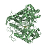
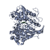
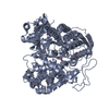
| Title | Structure of ligand free human DPP3 in closed form. | |||||||||
 Components Components | Dipeptidyl peptidase 3 | |||||||||
 Keywords Keywords | HYDROLASE / Unbound / Peptidase / Zinc | |||||||||
| Function / homology |  Function and homology information Function and homology informationdipeptidyl-peptidase III / metalloexopeptidase activity / dipeptidyl-peptidase activity / aminopeptidase activity / KEAP1-NFE2L2 pathway / Neddylation / proteolysis / extracellular exosome / zinc ion binding / cytosol / cytoplasm Similarity search - Function | |||||||||
| Biological species |  Homo sapiens (human) Homo sapiens (human) | |||||||||
| Method |  X-RAY DIFFRACTION / SYNCHROTRON / MOLECULAR REPLACEMENT / Resolution: 2.741 Å X-RAY DIFFRACTION / SYNCHROTRON / MOLECULAR REPLACEMENT / Resolution: 2.741 Å | |||||||||
 Authors Authors | Kumar, P. / Reithofer, V. / Reisinger, M. / Pavkov-Keller, T. / Wallner, S. / Macheroux, P. / Gruber, K. | |||||||||
| Funding support |  Austria, 1items Austria, 1items
| |||||||||
 Citation Citation | Journal: Sci Rep / Year: 2016 Title: Substrate complexes of human dipeptidyl peptidase III reveal the mechanism of enzyme inhibition. Authors: Kumar, P. / Reithofer, V. / Reisinger, M. / Wallner, S. / Pavkov-Keller, T. / Macheroux, P. / Gruber, K. | |||||||||
| History |
|
- Structure visualization
| Structure viewer | Molecule: MolmilJmol/JSmol |
|---|
- Downloads & links
-Download
| PDBx/mmCIF format | 5egy.cif.gz | 154.9 KB | Display | PDBx/mmCIF format |
|---|---|---|---|---|
| PDB format | pdb5egy.ent.gz | 121.9 KB | Display | PDB format |
| PDBx/mmJSON format | 5egy.json.gz | Tree view | PDBx/mmJSON format | |
| Others |  Other downloads Other downloads |
-Validation report
| Arichive directory | https://data.pdbj.org/pub/pdb/validation_reports/eg/5egyftp://data.pdbj.org/pub/pdb/validation_reports/eg/5egy | HTTPS FTP |
|---|
-Related structure data
| Related structure data | 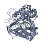 5e2qC  5e33C  5e3aC  5e3cC  5ehhC  3t6bS S: Starting model for refinement C: citing same article ( |
|---|---|
| Similar structure data |


-Links
 PDBj
PDBj
- Assembly
| Deposited unit | 
| ||||||||
|---|---|---|---|---|---|---|---|---|---|
| 1 |
| ||||||||
| Unit cell |
| ||||||||
| Components on special symmetry positions |
|
-Components
| #1: Protein | Mass: 81548.688 Da / Num. of mol.: 1 / Mutation: C19S, E207C, E451A, S491C, C519S, C654S Source method: isolated from a genetically manipulated source Source: (gene. exp.) Homo sapiens (human) / Gene: DPP3 / Plasmid: PET28MHL / Production host:  |
|---|---|
| #2: Chemical | ChemComp-ZN /   Mass: 65.409 Da / Num. of mol.: 1 / Source method: obtained synthetically / Formula: Zn Mass: 65.409 Da / Num. of mol.: 1 / Source method: obtained synthetically / Formula: Zn |
| #3: Chemical | ChemComp-MG /   Mass: 24.305 Da / Num. of mol.: 1 / Source method: obtained synthetically / Formula: Mg Mass: 24.305 Da / Num. of mol.: 1 / Source method: obtained synthetically / Formula: Mg |
| #4: Water | ChemComp-HOH /  Mass: 18.015 Da / Num. of mol.: 22 / Source method: isolated from a natural source / Formula: H2O Mass: 18.015 Da / Num. of mol.: 22 / Source method: isolated from a natural source / Formula: H2O |
| Has protein modification | Y |
-Experimental details
-Experiment
| Experiment | Method: X-RAY DIFFRACTION |
|---|
- Sample preparation
| Crystal | Density Matthews: 2.41 Å3/Da / Density % sol: 49.04 % |
|---|---|
| Crystal grow | Temperature: 293 K / Method: vapor diffusion, sitting drop / pH: 8.2 Details: 0.056 M Sodium phosphate monobasic monohydrate, 1.344M Potassium phosphate dibasic, pH- 8.2, 293K PH range: 8.2 |
-Data collection
| Diffraction | Mean temperature: 100 K |
|---|---|
| Diffraction source | Source: SYNCHROTRON / Site: ESRF  / Beamline: ID23-1 / Wavelength: 0.9789 Å / Beamline: ID23-1 / Wavelength: 0.9789 Å |
| Detector | Type: DECTRIS PILATUS 6M / Detector: PIXEL / Date: Mar 1, 2015 |
| Radiation | Protocol: SINGLE WAVELENGTH / Monochromatic (M) / Laue (L): M / Scattering type: x-ray |
| Radiation wavelength | Wavelength: 0.9789 Å / Relative weight: 1 |
| Reflection | Resolution: 2.74→48.83 Å / Num. all: 20281 / Num. obs: 20281 / % possible obs: 98.8 % / Redundancy: 3.3 % / Biso Wilson estimate: 69.01 Å2 / Rmerge(I) obs: 0.0723 / Rsym value: 0.0723 / Net I/σ(I): 12.01 |
| Reflection shell | Resolution: 2.74→2.83 Å / Redundancy: 3.2 % / Rmerge(I) obs: 0.7944 / Mean I/σ(I) obs: 1.64 / % possible all: 96.02 |
- Processing
| Software |
| ||||||||||||||||||||||||||||||||||||||||||||||||||||||||
|---|---|---|---|---|---|---|---|---|---|---|---|---|---|---|---|---|---|---|---|---|---|---|---|---|---|---|---|---|---|---|---|---|---|---|---|---|---|---|---|---|---|---|---|---|---|---|---|---|---|---|---|---|---|---|---|---|---|
| Refinement | Method to determine structure: MOLECULAR REPLACEMENT Starting model: 3T6B Resolution: 2.741→43.83 Å / SU ML: 0.41 / Cross valid method: FREE R-VALUE / σ(F): 1.36 / Phase error: 35 / Stereochemistry target values: ML
| ||||||||||||||||||||||||||||||||||||||||||||||||||||||||
| Solvent computation | Shrinkage radii: 0.9 Å / VDW probe radii: 1.11 Å / Solvent model: FLAT BULK SOLVENT MODEL | ||||||||||||||||||||||||||||||||||||||||||||||||||||||||
| Refinement step | Cycle: LAST / Resolution: 2.741→43.83 Å
| ||||||||||||||||||||||||||||||||||||||||||||||||||||||||
| Refine LS restraints |
| ||||||||||||||||||||||||||||||||||||||||||||||||||||||||
| LS refinement shell |
|
