 Movie
Movie Controller

[English] 日本語
 Yorodumi
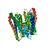
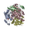
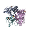
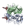
- PDB-1yrh: Crystal Structure Of Trp Repressor Binding Protein Wrba in comple... -
+ Open data

- Basic information
| Entry | Database: PDB / ID: 1yrh | ||||||
|---|---|---|---|---|---|---|---|
| Title | Crystal Structure Of Trp Repressor Binding Protein Wrba in complex with FMN | ||||||
 Components Components | trp repressor binding protein WrbA | ||||||
 Keywords Keywords | PROTEIN BINDING / alpha-beta twisted open sheet / Structural Genomics / PSI / Protein Structure Initiative / New York SGX Research Center for Structural Genomics / NYSGXRC | ||||||
| Function / homology |  Function and homology information Function and homology informationNAD(P)H dehydrogenase (quinone) / NADPH dehydrogenase (quinone) activity / NADH dehydrogenase (quinone) (non-electrogenic) activity / NAD(P)H dehydrogenase (quinone) activity / FMN binding / membrane Similarity search - Function | ||||||
| Biological species |  Deinococcus radiodurans (radioresistant) Deinococcus radiodurans (radioresistant) | ||||||
| Method |  X-RAY DIFFRACTION / SYNCHROTRON / SAD / Resolution: 3.11 Å X-RAY DIFFRACTION / SYNCHROTRON / SAD / Resolution: 3.11 Å | ||||||
 Authors Authors | Gorman, J. / Shapiro, L. / Burley, S.K. / New York SGX Research Center for Structural Genomics (NYSGXRC) | ||||||
 Citation Citation | Journal: Protein Sci. / Year: 2005 Title: Crystal structures of the tryptophan repressor binding protein WrbA and complexes with flavin mononucleotide. Authors: Gorman, J. / Shapiro, L. | ||||||
| History |
|
- Structure visualization
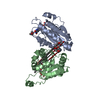
| Structure viewer | Molecule: MolmilJmol/JSmol |
|---|
- Downloads & links
-Download
| PDBx/mmCIF format | 1yrh.cif.gz | 305.4 KB | Display | PDBx/mmCIF format |
|---|---|---|---|---|
| PDB format | pdb1yrh.ent.gz | 250.8 KB | Display | PDB format |
| PDBx/mmJSON format | 1yrh.json.gz | Tree view | PDBx/mmJSON format | |
| Others |  Other downloads Other downloads |
-Validation report
| Arichive directory | https://data.pdbj.org/pub/pdb/validation_reports/yr/1yrhftp://data.pdbj.org/pub/pdb/validation_reports/yr/1yrh | HTTPS FTP |
|---|
-Related structure data
| Related structure data |  1ydgC  1zwkC  1zwlC C: citing same article ( |
|---|---|
| Similar structure data | |
| Other databases |


-Links
 PDBj
PDBj

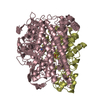
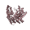
- Assembly
| Deposited unit | 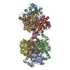
| ||||||||
|---|---|---|---|---|---|---|---|---|---|
| 1 | 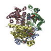
| ||||||||
| 2 | 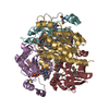
| ||||||||
| Unit cell |
| ||||||||
| Details | The biological assembly is a tetramer. There are two in the asymmetric unit: A, B, C, D form one biological unit and E, F, G, H form a second |
-Components
| #1: Protein | Mass: 22900.686 Da / Num. of mol.: 8 Source method: isolated from a genetically manipulated source Source: (gene. exp.) Deinococcus radiodurans (radioresistant)Gene: DRA0214 / Species (production host): Escherichia coli / Production host: #2: Chemical | ChemComp-FMN /   Mass: 456.344 Da / Num. of mol.: 8 / Source method: obtained synthetically / Formula: C17H21N4O9P Mass: 456.344 Da / Num. of mol.: 8 / Source method: obtained synthetically / Formula: C17H21N4O9P#3: Water | ChemComp-HOH / |  Mass: 18.015 Da / Num. of mol.: 161 / Source method: isolated from a natural source / Formula: H2O Mass: 18.015 Da / Num. of mol.: 161 / Source method: isolated from a natural source / Formula: H2OHas protein modification | Y | |
|---|
-Experimental details
-Experiment
| Experiment | Method: X-RAY DIFFRACTION / Number of used crystals: 1 |
|---|
- Sample preparation
| Crystal | Density Matthews: 2.6 Å3/Da / Density % sol: 52.9 % |
|---|---|
| Crystal grow | Temperature: 293 K / Method: vapor diffusion / pH: 4.5 Details: 2.6M AMSULFATE, 20% GLYCEROL, pH 4.5, VAPOR DIFFUSION, temperature 293K |
-Data collection
| Diffraction | Mean temperature: 100 K |
|---|---|
| Diffraction source | Source: SYNCHROTRON / Site: APS  / Beamline: 31-ID / Wavelength: 0.9793 Å / Beamline: 31-ID / Wavelength: 0.9793 Å |
| Detector | Type: MARRESEARCH / Detector: CCD / Date: Aug 9, 2004 |
| Radiation | Monochromator: DIAMOND / Protocol: SINGLE WAVELENGTH / Monochromatic (M) / Laue (L): M / Scattering type: x-ray |
| Radiation wavelength | Wavelength: 0.9793 Å / Relative weight: 1 |
| Reflection | Resolution: 3.1→20 Å / Num. all: 32107 / Num. obs: 32107 / Observed criterion σ(F): 0 / Observed criterion σ(I): -3 / Redundancy: 3.8 % / Biso Wilson estimate: 60.5 Å2 / Rmerge(I) obs: 0.17 / Net I/σ(I): 5.6 |
| Reflection shell | Resolution: 3.1→3.21 Å / Rmerge(I) obs: 0.32 / Mean I/σ(I) obs: 4.5 / Num. unique all: 3169 / % possible all: 97 |
- Processing
| Software |
| ||||||||||||||||||||||||||||||||||||||||||||||||||||||||||||||||||||||||||||||||||||||||||||||||||||||||||||||||||||||||||||||||||
|---|---|---|---|---|---|---|---|---|---|---|---|---|---|---|---|---|---|---|---|---|---|---|---|---|---|---|---|---|---|---|---|---|---|---|---|---|---|---|---|---|---|---|---|---|---|---|---|---|---|---|---|---|---|---|---|---|---|---|---|---|---|---|---|---|---|---|---|---|---|---|---|---|---|---|---|---|---|---|---|---|---|---|---|---|---|---|---|---|---|---|---|---|---|---|---|---|---|---|---|---|---|---|---|---|---|---|---|---|---|---|---|---|---|---|---|---|---|---|---|---|---|---|---|---|---|---|---|---|---|---|---|
| Refinement | Method to determine structure: SAD / Resolution: 3.11→20 Å / Cor.coef. Fo:Fc: 0.904 / Cor.coef. Fo:Fc free: 0.868 / SU B: 16.514 / SU ML: 0.282 / Cross valid method: THROUGHOUT / σ(F): 0 / σ(I): -3 / ESU R Free: 0.476 / Stereochemistry target values: MAXIMUM LIKELIHOOD / Details: HYDROGENS HAVE BEEN ADDED IN THE RIDING POSITIONS
| ||||||||||||||||||||||||||||||||||||||||||||||||||||||||||||||||||||||||||||||||||||||||||||||||||||||||||||||||||||||||||||||||||
| Solvent computation | Ion probe radii: 0.8 Å / Shrinkage radii: 0.8 Å / VDW probe radii: 1.2 Å / Solvent model: MASK | ||||||||||||||||||||||||||||||||||||||||||||||||||||||||||||||||||||||||||||||||||||||||||||||||||||||||||||||||||||||||||||||||||
| Displacement parameters | Biso mean: 32.47 Å2
| ||||||||||||||||||||||||||||||||||||||||||||||||||||||||||||||||||||||||||||||||||||||||||||||||||||||||||||||||||||||||||||||||||
| Refinement step | Cycle: LAST / Resolution: 3.11→20 Å
| ||||||||||||||||||||||||||||||||||||||||||||||||||||||||||||||||||||||||||||||||||||||||||||||||||||||||||||||||||||||||||||||||||
| Refine LS restraints |
| ||||||||||||||||||||||||||||||||||||||||||||||||||||||||||||||||||||||||||||||||||||||||||||||||||||||||||||||||||||||||||||||||||
| LS refinement shell | Resolution: 3.11→3.189 Å / Total num. of bins used: 20
|
