Protocol: SINGLE WAVELENGTH / Monochromatic (M) / Laue (L): M / Scattering type: x-ray
Radiation wavelength
Wavelength: 0.933 Å / Relative weight: 1
Reflection
Resolution: 2→47.6 Å / Num. obs: 117179 / % possible obs: 99.8 % / Redundancy: 3.63 % / Rmerge(I) obs: 0.01 / Net I/σ(I): 8.99
Reflection shell
Resolution: 2→2.1 Å / Redundancy: 3.61 % / Rmerge(I) obs: 0.05 / Mean I/σ(I) obs: 3.19 / % possible all: 100
-
Processing
Software
Name
Version
Classification
XDS
datareduction
SHELXD
phasing
SHARP
phasing
REFMAC
5.2.0019
refinement
Refinement
Method to determine structure: SAD Starting model: NONE Resolution: 2→47.57 Å / Cor.coef. Fo:Fc: 0.952 / Cor.coef. Fo:Fc free: 0.931 / SU B: 3.453 / SU ML: 0.095 / Cross valid method: THROUGHOUT / ESU R: 0.139 / ESU R Free: 0.134 / Stereochemistry target values: MAXIMUM LIKELIHOOD / Details: HYDROGENS HAVE BEEN ADDED IN THE RIDING POSITIONS
Rfactor
Num. reflection
% reflection
Selection details
Rfree
0.214
5865
5 %
RANDOM
Rwork
0.175
-
-
-
obs
0.177
111098
99.6 %
-
Solvent computation
Ion probe radii: 0.8 Å / Shrinkage radii: 0.8 Å / VDW probe radii: 1.4 Å / Solvent model: MASK
 Movie
Movie Controller

 Yorodumi
 Open data
 Basic information
 Components
 Keywords
 Function and homology information
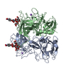
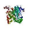
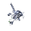
 FOWL ADENOVIRUS 1
 X-RAY DIFFRACTION /
  Authors
 Citation
 Structure visualization
 Downloads & links
 Other downloads
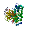
 PDBj
PDBj
 Assembly


 Mass: 96.063 Da / Num. of mol.: 10 / Source method: obtained synthetically / Formula: SO4
 Mass: 92.094 Da / Num. of mol.: 2 / Source method: obtained synthetically / Formula: C3H8O3
 Mass: 18.015 Da / Num. of mol.: 1177 / Source method: isolated from a natural source / Formula: H2O
 Sample preparation
 / Beamline: ID14-4 / Wavelength: 0.933
  Processing