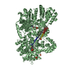
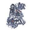
 Movie
Movie Controller


+ Open data

- Basic information
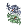
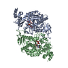
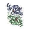
| Entry | Database: PDB / ID: 1jvd | ||||||
|---|---|---|---|---|---|---|---|
| Title | CRYSTAL STRUCTURE OF HUMAN AGX2 COMPLEXED WITH UDPGLCNAC | ||||||
 Components Components | UDPGLCNAC PYROPHOSPHORYLASE (AGX2) | ||||||
 Keywords Keywords | TRANSFERASE / NUCLEOTIDYLTRANSFERASE / ALTERNATIVE SPLICING | ||||||
| Function / homology |  Function and homology information Function and homology informationprotein serine pyrophosphorylase activity / UDP-N-acetylgalactosamine diphosphorylase / UDP-N-acetylgalactosamine diphosphorylase activity / Transferases; Transferring phosphorus-containing groups; Phosphotransferases with a phosphate group as acceptor / Synthesis of UDP-N-acetyl-glucosamine / UDP-N-acetylglucosamine diphosphorylase / UDP-N-acetylglucosamine diphosphorylase activity / UDP-N-acetylglucosamine biosynthetic process / positive regulation of type I interferon production / antiviral innate immune response ...protein serine pyrophosphorylase activity / UDP-N-acetylgalactosamine diphosphorylase / UDP-N-acetylgalactosamine diphosphorylase activity / Transferases; Transferring phosphorus-containing groups; Phosphotransferases with a phosphate group as acceptor / Synthesis of UDP-N-acetyl-glucosamine / UDP-N-acetylglucosamine diphosphorylase / UDP-N-acetylglucosamine diphosphorylase activity / UDP-N-acetylglucosamine biosynthetic process / positive regulation of type I interferon production / antiviral innate immune response / nucleoplasm / identical protein binding / plasma membrane / cytosol Similarity search - Function | ||||||
| Biological species |  Homo sapiens (human) Homo sapiens (human) | ||||||
| Method |  X-RAY DIFFRACTION / SYNCHROTRON / MOLECULAR REPLACEMENT / Resolution: 2.4 Å X-RAY DIFFRACTION / SYNCHROTRON / MOLECULAR REPLACEMENT / Resolution: 2.4 Å | ||||||
 Authors Authors | Peneff, C. / Bourne, Y. | ||||||
 Citation Citation | Journal: EMBO J. / Year: 2001 Title: Crystal structures of two human pyrophosphorylase isoforms in complexes with UDPGlc(Gal)NAc: role of the alternatively spliced insert in the enzyme oligomeric assembly and active site architecture. Authors: Peneff, C. / Ferrari, P. / Charrier, V. / Taburet, Y. / Monnier, C. / Zamboni, V. / Winter, J. / Harnois, M. / Fassy, F. / Bourne, Y. | ||||||
| History |
|
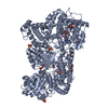
- Structure visualization
| Structure viewer | Molecule: MolmilJmol/JSmol |
|---|
- Downloads & links
-Download
| PDBx/mmCIF format | 1jvd.cif.gz | 209.2 KB | Display | PDBx/mmCIF format |
|---|---|---|---|---|
| PDB format | pdb1jvd.ent.gz | 165.3 KB | Display | PDB format |
| PDBx/mmJSON format | 1jvd.json.gz | Tree view | PDBx/mmJSON format | |
| Others |  Other downloads Other downloads |
-Validation report
| Arichive directory | https://data.pdbj.org/pub/pdb/validation_reports/jv/1jvdftp://data.pdbj.org/pub/pdb/validation_reports/jv/1jvd | HTTPS FTP |
|---|
-Related structure data


-Links
 PDBj
PDBj

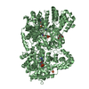
- Assembly
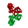
| Deposited unit | 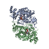
| ||||||||
|---|---|---|---|---|---|---|---|---|---|
| 1 |
| ||||||||
| Unit cell |
|
-Components
| #1: Protein | Mass: 58838.785 Da / Num. of mol.: 2 Source method: isolated from a genetically manipulated source Source: (gene. exp.) Homo sapiens (human) / Gene: AGX2 / Plasmid: pRU277 / Production host:  References: UniProt: Q16222, UDP-N-acetylglucosamine diphosphorylase #2: Chemical |   Mass: 607.354 Da / Num. of mol.: 2 / Source method: obtained synthetically / Formula: C17H27N3O17P2 Mass: 607.354 Da / Num. of mol.: 2 / Source method: obtained synthetically / Formula: C17H27N3O17P2#3: Water | ChemComp-HOH / |  Mass: 18.015 Da / Num. of mol.: 310 / Source method: isolated from a natural source / Formula: H2O Mass: 18.015 Da / Num. of mol.: 310 / Source method: isolated from a natural source / Formula: H2O |
|---|
-Experimental details
-Experiment
| Experiment | Method: X-RAY DIFFRACTION / Number of used crystals: 1 |
|---|
- Sample preparation
| Crystal | Density Matthews: 2.39 Å3/Da / Density % sol: 48.57 % | |||||||||||||||||||||||||||||||||||||||||||||||||
|---|---|---|---|---|---|---|---|---|---|---|---|---|---|---|---|---|---|---|---|---|---|---|---|---|---|---|---|---|---|---|---|---|---|---|---|---|---|---|---|---|---|---|---|---|---|---|---|---|---|---|
| Crystal grow | Temperature: 293 K / Method: vapor diffusion, hanging drop / pH: 5.7 Details: PEG600, pH 5.7, VAPOR DIFFUSION, HANGING DROP, temperature 293K | |||||||||||||||||||||||||||||||||||||||||||||||||
| Crystal grow | *PLUS Temperature: 20 ℃ | |||||||||||||||||||||||||||||||||||||||||||||||||
| Components of the solutions | *PLUS
|
-Data collection
| Diffraction | Mean temperature: 100 K |
|---|---|
| Diffraction source | Source: SYNCHROTRON / Site: ESRF  / Beamline: ID14-3 / Wavelength: 0.931 Å / Beamline: ID14-3 / Wavelength: 0.931 Å |
| Detector | Type: MARRESEARCH / Detector: CCD / Date: May 20, 1999 |
| Radiation | Protocol: SINGLE WAVELENGTH / Monochromatic (M) / Laue (L): M / Scattering type: x-ray |
| Radiation wavelength | Wavelength: 0.931 Å / Relative weight: 1 |
| Reflection | Resolution: 2.4→40 Å / Num. obs: 43625 / % possible obs: 99.8 % / Observed criterion σ(I): 2 / Redundancy: 3.5 % / Biso Wilson estimate: 35 Å2 / Rsym value: 0.062 / Net I/σ(I): 6.2 |
| Reflection | *PLUS Lowest resolution: 40 Å / Rmerge(I) obs: 0.062 |
| Reflection shell | *PLUS % possible obs: 99.8 % / Rmerge(I) obs: 0.273 / Mean I/σ(I) obs: 2.6 |
- Processing
| Software |
| ||||||||||||||||||||||||||||||||||||||||||||||||||||||||||||
|---|---|---|---|---|---|---|---|---|---|---|---|---|---|---|---|---|---|---|---|---|---|---|---|---|---|---|---|---|---|---|---|---|---|---|---|---|---|---|---|---|---|---|---|---|---|---|---|---|---|---|---|---|---|---|---|---|---|---|---|---|---|
| Refinement | Method to determine structure: MOLECULAR REPLACEMENT / Resolution: 2.4→19.97 Å / Rfactor Rfree error: 0.005 / Data cutoff high absF: 1498298.17 / Data cutoff low absF: 0 / Isotropic thermal model: RESTRAINED / Cross valid method: THROUGHOUT / σ(F): 0 / Stereochemistry target values: Engh & Huber
| ||||||||||||||||||||||||||||||||||||||||||||||||||||||||||||
| Solvent computation | Solvent model: FLAT MODEL / Bsol: 40.6024 Å2 / ksol: 0.353303 e/Å3 | ||||||||||||||||||||||||||||||||||||||||||||||||||||||||||||
| Displacement parameters | Biso mean: 38.6 Å2
| ||||||||||||||||||||||||||||||||||||||||||||||||||||||||||||
| Refine analyze |
| ||||||||||||||||||||||||||||||||||||||||||||||||||||||||||||
| Refinement step | Cycle: LAST / Resolution: 2.4→19.97 Å
| ||||||||||||||||||||||||||||||||||||||||||||||||||||||||||||
| Refine LS restraints |
| ||||||||||||||||||||||||||||||||||||||||||||||||||||||||||||
| LS refinement shell | Resolution: 2.4→2.55 Å / Rfactor Rfree error: 0.017 / Total num. of bins used: 6
| ||||||||||||||||||||||||||||||||||||||||||||||||||||||||||||
| Xplor file |
| ||||||||||||||||||||||||||||||||||||||||||||||||||||||||||||
| Refinement | *PLUS Highest resolution: 2.4 Å / Lowest resolution: 20 Å / Rfactor Rfree: 0.251 / Rfactor Rwork: 0.193 | ||||||||||||||||||||||||||||||||||||||||||||||||||||||||||||
| Solvent computation | *PLUS | ||||||||||||||||||||||||||||||||||||||||||||||||||||||||||||
| Displacement parameters | *PLUS | ||||||||||||||||||||||||||||||||||||||||||||||||||||||||||||
| Refine LS restraints | *PLUS
| ||||||||||||||||||||||||||||||||||||||||||||||||||||||||||||
| LS refinement shell | *PLUS Rfactor Rfree: 0.309 / Rfactor Rwork: 0.242 |
