 Movie
Movie Controller


+ Open data

- Basic information
| Entry | Database: PDB / ID: 4yir | ||||||||||||
|---|---|---|---|---|---|---|---|---|---|---|---|---|---|
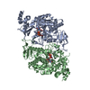
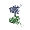
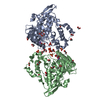
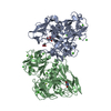
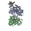
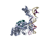
| Title | Crystal structure of Rad4-Rad23 crosslinked to an undamaged DNA | ||||||||||||
 Components Components |
| ||||||||||||
 Keywords Keywords | DNA BINDING PROTEIN/DNA / DNA damage repair / nucleotide excision repair / protein-DNA interactions / protein-DNA crosslinking / protein-DNA complex / xeroderma pigmentosum / beta-hairpin / transglutaminase domain / disulfide crosslinking / DNA BINDING PROTEIN-DNA complex | ||||||||||||
| Function / homology |  Function and homology information Function and homology informationPNGase complex / single-strand break-containing DNA binding / nucleotide-excision repair factor 2 complex / XPC complex / nucleotide-excision repair, DNA damage recognition / K48-linked polyubiquitin modification-dependent protein binding / proteasome binding / DNA topological change / polyubiquitin modification-dependent protein binding / mismatch repair ...PNGase complex / single-strand break-containing DNA binding / nucleotide-excision repair factor 2 complex / XPC complex / nucleotide-excision repair, DNA damage recognition / K48-linked polyubiquitin modification-dependent protein binding / proteasome binding / DNA topological change / polyubiquitin modification-dependent protein binding / mismatch repair / ERAD pathway / nucleotide-excision repair / ubiquitin binding / single-stranded DNA binding / damaged DNA binding / proteasome-mediated ubiquitin-dependent protein catabolic process / protein-macromolecule adaptor activity / negative regulation of transcription by RNA polymerase II / mitochondrion / nucleoplasm / nucleus / cytoplasm / cytosol Similarity search - Function | ||||||||||||
| Biological species |  synthetic construct (others) | ||||||||||||
| Method |  X-RAY DIFFRACTION / SYNCHROTRON / MOLECULAR REPLACEMENT / Resolution: 3.0501 Å X-RAY DIFFRACTION / SYNCHROTRON / MOLECULAR REPLACEMENT / Resolution: 3.0501 Å | ||||||||||||
 Authors Authors | Min, J.-H. / Chen, X. / Kim, Y. | ||||||||||||
| Funding support |  United States, 3items United States, 3items
| ||||||||||||
 Citation Citation | Journal: Nat Commun / Year: 2015 Title: Kinetic gating mechanism of DNA damage recognition by Rad4/XPC. Authors: Chen, X. / Velmurugu, Y. / Zheng, G. / Park, B. / Shim, Y. / Kim, Y. / Liu, L. / Van Houten, B. / He, C. / Ansari, A. / Min, J.H. | ||||||||||||
| History |
|
- Structure visualization
| Structure viewer | Molecule: MolmilJmol/JSmol |
|---|
- Downloads & links
-Download
| PDBx/mmCIF format | 4yir.cif.gz | 406.8 KB | Display | PDBx/mmCIF format |
|---|---|---|---|---|
| PDB format | pdb4yir.ent.gz | 333.4 KB | Display | PDB format |
| PDBx/mmJSON format | 4yir.json.gz | Tree view | PDBx/mmJSON format | |
| Others |  Other downloads Other downloads |
-Validation report
| Arichive directory | https://data.pdbj.org/pub/pdb/validation_reports/yi/4yirftp://data.pdbj.org/pub/pdb/validation_reports/yi/4yir | HTTPS FTP |
|---|
-Related structure data
| Related structure data |  6ubfC  2qshS S: Starting model for refinement C: citing same article ( |
|---|---|
| Similar structure data |


-Links
 PDBj
PDBj


- Assembly
| Deposited unit | 
| ||||||||
|---|---|---|---|---|---|---|---|---|---|
| 1 |
| ||||||||
| Unit cell |
|
-Components
| #1: Protein | Mass: 62588.508 Da / Num. of mol.: 1 Source method: isolated from a genetically manipulated source Source: (gene. exp.) Strain: ATCC 204508 / S288c / Gene: RAD4, YER162C / Cell line (production host): High Five Cells (BTI-TN-5B1-4) / Production host:  Trichoplusia ni (cabbage looper) / References: UniProt: P14736 Trichoplusia ni (cabbage looper) / References: UniProt: P14736 |
|---|---|
| #2: Protein | Mass: 17783.352 Da / Num. of mol.: 1 Source method: isolated from a genetically manipulated source Source: (gene. exp.) Strain: ATCC 204508 / S288c / Gene: RAD23, YEL037C, SYGP-ORF29 / Cell line (production host): High Five Cells (BTI-TN-5B1-4) / Production host: Trichoplusia ni (cabbage looper) / References: UniProt: P32628 |
| #3: DNA chain | Mass: 7294.800 Da / Num. of mol.: 1 Source method: isolated from a genetically manipulated source Source: (gene. exp.) synthetic construct (others) / Production host: synthetic construct (others) |
| #4: DNA chain | Mass: 7505.837 Da / Num. of mol.: 1 Source method: isolated from a genetically manipulated source Source: (gene. exp.) synthetic construct (others) / Production host: synthetic construct (others) |
| Has protein modification | Y |
-Experimental details
-Experiment
| Experiment | Method: X-RAY DIFFRACTION / Number of used crystals: 1 |
|---|
- Sample preparation
| Crystal | Density Matthews: 3.35 Å3/Da / Density % sol: 63.27 % |
|---|---|
| Crystal grow | Temperature: 277 K / Method: vapor diffusion, hanging drop Details: 50mM BTP-HCl, 100mM NaCl, 14% isopropanol and 100mM calcium chloride PH range: 6.8 |
-Data collection
| Diffraction | Mean temperature: 103 K |
|---|---|
| Diffraction source | Source: SYNCHROTRON / Site: APS / Beamline: 21-ID-D / Wavelength: 0.97919 Å |
| Detector | Type: MARMOSAIC 300 mm CCD / Detector: CCD / Date: Apr 18, 2013 |
| Radiation | Protocol: SINGLE WAVELENGTH / Monochromatic (M) / Laue (L): M / Scattering type: x-ray |
| Radiation wavelength | Wavelength: 0.97919 Å / Relative weight: 1 |
| Reflection | Resolution: 3.05→39.7 Å / Num. obs: 45912 / % possible obs: 97.51 % / Redundancy: 6.7 % / Rmerge(I) obs: 0.071 / Net I/σ(I): 23.8 |
| Reflection shell | Resolution: 3.05→3.1 Å / Redundancy: 6.9 % / Rmerge(I) obs: 0.792 / Num. unique all: 1228 / % possible all: 100 |
- Processing
| Software |
| ||||||||||||||||||||||||||||||||||||||||||||||||||||||||||||||||||||||||||||||||||||||||||||||||||||||||||||||||||||||||||||||||||||||||||||||||||||||||||||||||||||||||||||||||||||||
|---|---|---|---|---|---|---|---|---|---|---|---|---|---|---|---|---|---|---|---|---|---|---|---|---|---|---|---|---|---|---|---|---|---|---|---|---|---|---|---|---|---|---|---|---|---|---|---|---|---|---|---|---|---|---|---|---|---|---|---|---|---|---|---|---|---|---|---|---|---|---|---|---|---|---|---|---|---|---|---|---|---|---|---|---|---|---|---|---|---|---|---|---|---|---|---|---|---|---|---|---|---|---|---|---|---|---|---|---|---|---|---|---|---|---|---|---|---|---|---|---|---|---|---|---|---|---|---|---|---|---|---|---|---|---|---|---|---|---|---|---|---|---|---|---|---|---|---|---|---|---|---|---|---|---|---|---|---|---|---|---|---|---|---|---|---|---|---|---|---|---|---|---|---|---|---|---|---|---|---|---|---|---|---|
| Refinement | Method to determine structure: MOLECULAR REPLACEMENT Starting model: 2QSH Resolution: 3.0501→39.7 Å / SU ML: 0.37 / Cross valid method: FREE R-VALUE / σ(F): 0.18 / Phase error: 24.88 / Stereochemistry target values: ML
| ||||||||||||||||||||||||||||||||||||||||||||||||||||||||||||||||||||||||||||||||||||||||||||||||||||||||||||||||||||||||||||||||||||||||||||||||||||||||||||||||||||||||||||||||||||||
| Solvent computation | Shrinkage radii: 0.9 Å / VDW probe radii: 1.11 Å / Solvent model: FLAT BULK SOLVENT MODEL | ||||||||||||||||||||||||||||||||||||||||||||||||||||||||||||||||||||||||||||||||||||||||||||||||||||||||||||||||||||||||||||||||||||||||||||||||||||||||||||||||||||||||||||||||||||||
| Refinement step | Cycle: LAST / Resolution: 3.0501→39.7 Å
| ||||||||||||||||||||||||||||||||||||||||||||||||||||||||||||||||||||||||||||||||||||||||||||||||||||||||||||||||||||||||||||||||||||||||||||||||||||||||||||||||||||||||||||||||||||||
| Refine LS restraints |
| ||||||||||||||||||||||||||||||||||||||||||||||||||||||||||||||||||||||||||||||||||||||||||||||||||||||||||||||||||||||||||||||||||||||||||||||||||||||||||||||||||||||||||||||||||||||
| LS refinement shell |
| ||||||||||||||||||||||||||||||||||||||||||||||||||||||||||||||||||||||||||||||||||||||||||||||||||||||||||||||||||||||||||||||||||||||||||||||||||||||||||||||||||||||||||||||||||||||
| Refinement TLS params. | Method: refined / Origin x: 35.4288 Å / Origin y: 28.4702 Å / Origin z: 437.0431 Å
| ||||||||||||||||||||||||||||||||||||||||||||||||||||||||||||||||||||||||||||||||||||||||||||||||||||||||||||||||||||||||||||||||||||||||||||||||||||||||||||||||||||||||||||||||||||||
| Refinement TLS group | Selection details: all |
