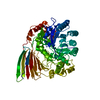
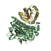
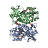
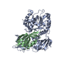
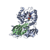
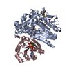
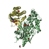
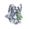
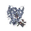
- PDB-6q2t: Human sterol 14a-demethylase (CYP51) in complex with the function... -
+
Open data
ID or keywords:
Loading...
-
Basic information
Entry
Database: PDB / ID: 6q2t
Title
Human sterol 14a-demethylase (CYP51) in complex with the functionally irreversible inhibitor (R)-N-(1-(3-chloro-4'-fluoro-[1,1'-biphenyl]-4-yl)-2-(1H-imidazol-1-yl)ethyl)-4-(5-(3-fluoro-5-(5-fluoropyrimidin-4-yl)phenyl)-1,3,4-oxadiazol-2-yl)benzamide
: / sterol 14alpha-demethylase / sterol 14-demethylase activity / negative regulation of amyloid-beta clearance / sterol metabolic process / steroid biosynthetic process / Cholesterol biosynthesis / EGR2 and SOX10-mediated initiation of Schwann cell myelination / oxidoreductase activity, acting on paired donors, with incorporation or reduction of molecular oxygen, reduced flavin or flavoprotein as one donor, and incorporation of one atom of oxygen / cholesterol biosynthetic process ...: / sterol 14alpha-demethylase / sterol 14-demethylase activity / negative regulation of amyloid-beta clearance / sterol metabolic process / steroid biosynthetic process / Cholesterol biosynthesis / EGR2 and SOX10-mediated initiation of Schwann cell myelination / oxidoreductase activity, acting on paired donors, with incorporation or reduction of molecular oxygen, reduced flavin or flavoprotein as one donor, and incorporation of one atom of oxygen / cholesterol biosynthetic process / negative regulation of protein secretion / Endogenous sterols / Activation of gene expression by SREBF (SREBP) / negative regulation of protein catabolic process / oxidoreductase activity / iron ion binding / heme binding / endoplasmic reticulum membrane / membrane Similarity search - Function
Resolution: 2.8→28.098 Å / Cor.coef. Fo:Fc: 0.955 / Cor.coef. Fo:Fc free: 0.937 / SU B: 23.112 / SU ML: 0.417 / Cross valid method: THROUGHOUT / ESU R Free: 0.403 Details: Hydrogens have been added in their riding positions
Rfactor
Num. reflection
% reflection
Rfree
0.2559
1258
4.785 %
Rwork
0.2268
-
-
all
0.228
-
-
obs
-
26289
99.561 %
Solvent computation
Ion probe radii: 0.8 Å / Shrinkage radii: 0.8 Å / VDW probe radii: 1.2 Å
Displacement parameters
Baniso -1
Baniso -2
Baniso -3
1-
1.581 Å2
-0 Å2
-0 Å2
2-
-
1.581 Å2
-0 Å2
3-
-
-
-3.162 Å2
Refinement step
Cycle: LAST / Resolution: 2.8→28.098 Å
Protein
Nucleic acid
Ligand
Solvent
Total
Num. atoms
7172
0
457
29
7658
Refine LS restraints
Refine-ID
Type
Dev ideal
Dev ideal target
Number
X-RAY DIFFRACTION
r_bond_refined_d
0.003
0.014
7871
X-RAY DIFFRACTION
r_bond_other_d
0.002
0.018
7284
X-RAY DIFFRACTION
r_angle_refined_deg
1.166
1.741
10757
X-RAY DIFFRACTION
r_angle_other_deg
1.134
1.676
16933
X-RAY DIFFRACTION
r_dihedral_angle_1_deg
6.165
5
887
X-RAY DIFFRACTION
r_dihedral_angle_2_deg
31.611
21.744
390
X-RAY DIFFRACTION
r_dihedral_angle_3_deg
14.007
15
1280
X-RAY DIFFRACTION
r_dihedral_angle_4_deg
13.019
15
50
X-RAY DIFFRACTION
r_chiral_restr
0.041
0.2
1023
X-RAY DIFFRACTION
r_chiral_restr_other
1.232
0.2
107
X-RAY DIFFRACTION
r_gen_planes_refined
0.007
0.02
8483
X-RAY DIFFRACTION
r_gen_planes_other
0.003
0.02
1699
X-RAY DIFFRACTION
r_nbd_refined
0.239
0.2
1933
X-RAY DIFFRACTION
r_symmetry_nbd_other
0.258
0.2
7508
X-RAY DIFFRACTION
r_nbtor_refined
0.178
0.2
3861
X-RAY DIFFRACTION
r_symmetry_nbtor_other
0.085
0.2
3133
X-RAY DIFFRACTION
r_xyhbond_nbd_refined
0.176
0.2
200
X-RAY DIFFRACTION
r_symmetry_xyhbond_nbd_other
0.083
0.2
6
X-RAY DIFFRACTION
r_symmetry_nbd_refined
0.298
0.2
20
X-RAY DIFFRACTION
r_nbd_other
0.351
0.2
112
X-RAY DIFFRACTION
r_symmetry_xyhbond_nbd_refined
0.275
0.2
3
X-RAY DIFFRACTION
r_mcbond_it
7.348
10.068
3554
X-RAY DIFFRACTION
r_mcbond_other
7.342
10.067
3553
X-RAY DIFFRACTION
r_mcangle_it
11.129
15.09
4439
X-RAY DIFFRACTION
r_mcangle_other
11.129
15.092
4440
X-RAY DIFFRACTION
r_scbond_it
6.923
10.414
4316
X-RAY DIFFRACTION
r_scbond_other
6.922
10.413
4317
X-RAY DIFFRACTION
r_scangle_it
10.792
15.366
6318
X-RAY DIFFRACTION
r_scangle_other
10.791
15.365
6319
X-RAY DIFFRACTION
r_lrange_it
15.389
117.991
9179
X-RAY DIFFRACTION
r_lrange_other
15.388
117.985
9180
LS refinement shell
Resolution (Å)
Rfactor Rfree
Num. reflection Rfree
Rfactor Rwork
Num. reflection Rwork
Refine-ID
% reflection obs (%)
2.8-2.873
0.387
77
0.374
1871
X-RAY DIFFRACTION
99.9487
2.873-2.952
0.365
84
0.375
1811
X-RAY DIFFRACTION
99.8946
2.952-3.037
0.38
83
0.355
1737
X-RAY DIFFRACTION
99.8902
3.037-3.13
0.392
94
0.341
1695
X-RAY DIFFRACTION
99.9441
3.13-3.233
0.323
79
0.322
1657
X-RAY DIFFRACTION
99.9424
3.233-3.346
0.345
72
0.297
1604
X-RAY DIFFRACTION
99.9404
3.346-3.472
0.328
68
0.277
1586
X-RAY DIFFRACTION
99.819
3.472-3.614
0.32
55
0.262
1481
X-RAY DIFFRACTION
99.7403
3.614-3.774
0.314
84
0.263
1381
X-RAY DIFFRACTION
99.6599
3.774-3.958
0.306
84
0.245
1387
X-RAY DIFFRACTION
99.6612
3.958-4.172
0.24
92
0.224
1238
X-RAY DIFFRACTION
99.5509
4.172-4.425
0.219
74
0.21
1213
X-RAY DIFFRACTION
99.536
4.425-4.729
0.258
61
0.188
1147
X-RAY DIFFRACTION
99.67
4.729-5.107
0.264
40
0.195
1087
X-RAY DIFFRACTION
99.4704
5.107-5.593
0.237
65
0.193
978
X-RAY DIFFRACTION
99.618
5.593-6.251
0.23
45
0.204
887
X-RAY DIFFRACTION
99.3603
6.251-7.212
0.176
37
0.194
802
X-RAY DIFFRACTION
99.1726
7.212-8.82
0.128
39
0.156
675
X-RAY DIFFRACTION
99.1667
8.82-12.42
0.165
18
0.143
521
X-RAY DIFFRACTION
98.7179
12.42-28
0.29
7
0.231
273
X-RAY DIFFRACTION
87.7743
+
About Yorodumi
-
News
-
Feb 9, 2022. New format data for meta-information of EMDB entries
New format data for meta-information of EMDB entries
Version 3 of the EMDB header file is now the official format.
The previous official version 1.9 will be removed from the archive.
In the structure databanks used in Yorodumi, some data are registered as the other names, "COVID-19 virus" and "2019-nCoV". Here are the details of the virus and the list of structure data.
Jan 31, 2019. EMDB accession codes are about to change! (news from PDBe EMDB page)
EMDB accession codes are about to change! (news from PDBe EMDB page)
The allocation of 4 digits for EMDB accession codes will soon come to an end. Whilst these codes will remain in use, new EMDB accession codes will include an additional digit and will expand incrementally as the available range of codes is exhausted. The current 4-digit format prefixed with “EMD-” (i.e. EMD-XXXX) will advance to a 5-digit format (i.e. EMD-XXXXX), and so on. It is currently estimated that the 4-digit codes will be depleted around Spring 2019, at which point the 5-digit format will come into force.
The EM Navigator/Yorodumi systems omit the EMD- prefix.
Related info.:Q: What is EMD? / ID/Accession-code notation in Yorodumi/EM Navigator
Yorodumi is a browser for structure data from EMDB, PDB, SASBDB, etc.
This page is also the successor to EM Navigator detail page, and also detail information page/front-end page for Omokage search.
The word "yorodu" (or yorozu) is an old Japanese word meaning "ten thousand". "mi" (miru) is to see.
Related info.:EMDB / PDB / SASBDB / Comparison of 3 databanks / Yorodumi Search / Aug 31, 2016. New EM Navigator & Yorodumi / Yorodumi Papers / Jmol/JSmol / Function and homology information / Changes in new EM Navigator and Yorodumi
 Movie
Movie Controller

 Yorodumi
 Open data
 Basic information
 Components
 Keywords
 Function and homology information
 Homo sapiens (human)
 X-RAY DIFFRACTION /
  Authors
 United States, 1items
  Citation
 Structure visualization
 Downloads & links
 Other downloads


 PDBj
PDBj


 Assembly
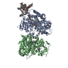


 Mass: 616.487 Da / Num. of mol.: 2 / Source method: obtained synthetically / Formula: C34H32FeN4O4
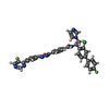
 Mass: 678.062 Da / Num. of mol.: 3 / Source method: obtained synthetically / Formula: C36H23ClF3N7O2 / Feature type: SUBJECT OF INVESTIGATION
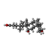
 Mass: 392.572 Da / Num. of mol.: 8 / Source method: obtained synthetically / Formula: C24H40O4 / Comment: detergent*YM
 Mass: 18.015 Da / Num. of mol.: 29 / Source method: isolated from a natural source / Formula: H2O
 Sample preparation
 Processing