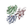
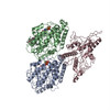
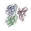
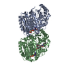
ジャーナル: J Mol Biol / 年: 2001 タイトル: Refined structure of alpha beta-tubulin at 3.5 A resolution. 著者: J Löwe / H Li / K H Downing / E Nogales / 要旨: We present a refined model of the alpha beta-tubulin dimer to 3.5 A resolution. An improved experimental density for the zinc-induced tubulin sheets was obtained by adding 114 electron diffraction ...We present a refined model of the alpha beta-tubulin dimer to 3.5 A resolution. An improved experimental density for the zinc-induced tubulin sheets was obtained by adding 114 electron diffraction patterns at 40-60 degrees tilt and increasing the completeness of structure factor amplitudes to 84.7 %. The refined structure was obtained using maximum-likelihood including phase information from experimental images, and simulated annealing Cartesian refinement to an R-factor of 23.2 and free R-factor of 29.7. The current model includes residues alpha:2-34, alpha:61-439, beta:2-437, one molecule of GTP, one of GDP, and one of taxol, as well as one magnesium ion at the non-exchangeable nucleotide site, and one putative zinc ion near the M-loop in the alpha-tubulin subunit. The acidic C-terminal tails could not be traced accurately, neither could the N-terminal loop including residues 35-60 in the alpha-subunit. There are no major changes in the overall fold of tubulin with respect to the previous structure, testifying to the quality of the initial experimental phases. The overall geometry of the model is, however, greatly improved, and the position of side-chains, especially those of exposed polar/charged groups, is much better defined. Three short protein sequence frame shifts were detected with respect to the non-refined structure. In light of the new model we discuss details of the tubulin structure such as nucleotide and taxol binding sites, lateral contacts in zinc-sheets, and the significance of the location of highly conserved residues.
THE SAMPLE WAS BOVINE, BUT THE MODELED PROTEIN SEQUENCES ARE FROM SUS SCROFA (PIG). THE AUTHORS ...THE SAMPLE WAS BOVINE, BUT THE MODELED PROTEIN SEQUENCES ARE FROM SUS SCROFA (PIG). THE AUTHORS USED THE SEQUENCES FROM THE MOST ABUNDANT ISOTYPE OF PIG BRAIN TUBULIN. THE RESIDUES IN CHAIN B ARE NOT SEQUENTIALLY NUMBERED. RESIDUES 44 AND 47 AND RESIDUES 360 AND 369 ARE COVALENTLY BOUND.
-
実験情報
-
実験
実験
手法: 電子線結晶学 / 使用した結晶の数: 200
EM実験
試料の集合状態: 2D ARRAY / 3次元再構成法: 電子線結晶学
-
試料調製
構成要素
名称: Alpha-Beta-tubulin sheets / タイプ: COMPLEX
試料
包埋: YES / シャドウイング: NO / 染色: NO / 凍結: YES
EM embedding
Material: tannin
急速凍結
凍結剤: NITROGEN
結晶
解説: JEOL 4000 electron microscope was used. Kodak film and gatan CCD were used as detectors. Temperature was 93 Kelvin.
結晶化
温度: 305 K / 手法: aberrant polymerization of tubulin / pH: 5.8 詳細: Zn++, pH 5.8, aberrant polymerization of tubulin, temperature 305K
結晶化
*PLUS
手法: unknown
-
データ収集
EM imaging
ID
照射モード
モデル
モード
Specimen-ID
1
SPOTSCAN
JEOL 4000
BRIGHTFIELD
1
2
FLOODBEAM
JEOL 4000
DIFFRACTION
1
撮影
フィルム・検出器のモデル: GENERIC FILM / 詳細: low dose
回折
ID
平均測定温度 (K)
Crystal-ID
1
93
1
2
1
放射光源
由来: ELECTRON MICROSCOPE / タイプ: JEOL 4000 electron microscope / 波長: 0.0194 Å
検出器
タイプ
ID
検出器
日付
KODAK
1
FILM
1994年1月1日
GATAN
2
CCD
放射
プロトコル: SINGLE WAVELENGTH / 単色(M)・ラウエ(L): M / 散乱光タイプ: electron
 ムービー
ムービー コントローラー


 データを開く
 基本情報
基本情報 要素
要素 キーワード
キーワード 機能・相同性情報
 分子置換 / クライオ電子顕微鏡法 / 解像度: 3.5 Å
  データ登録者
 引用
引用
 構造の表示
構造の表示 ダウンロードとリンク
 その他のダウンロード


 PDBj
PDBj


 集合体
集合体


 分子量: 65.409 Da / 分子数: 1 / 由来タイプ: 合成 / 式: Zn
 分子量: 24.305 Da / 分子数: 1 / 由来タイプ: 合成 / 式: Mg
 分子量: 523.180 Da / 分子数: 1 / 由来タイプ: 合成 / 式: C10H16N5O14P3 / コメント: GTP, エネルギー貯蔵分子*YM
 タイプ: RNA linking / 分子量: 443.201 Da / 分子数: 1 / 由来タイプ: 合成 / 式: C10H15N5O11P2 / コメント: GDP, エネルギー貯蔵分子*YM
 分子量: 853.906 Da / 分子数: 1 / 由来タイプ: 合成 / 式: C47H51NO14 / コメント: 薬剤*YM
 試料調製
試料調製 解析
解析