 Movie
Movie Controller


+ Open data

- Basic information
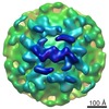
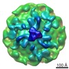
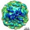
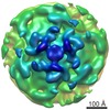
| Entry | Database: PDB / ID: 3edl | ||||||
|---|---|---|---|---|---|---|---|
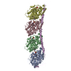
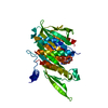
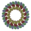
| Title | Kinesin13-Microtubule Ring complex | ||||||
 Components Components |
| ||||||
 Keywords Keywords | STRUCTURAL PROTEIN / Kinesin / Kinesin13 / Kin-I / M-Kinesin / Microtubule / Tubulin / depolymerization | ||||||
| Function / homology |  Function and homology information Function and homology informationpostsynaptic cytoskeleton organization / regulation of chromosome segregation / establishment or maintenance of microtubule cytoskeleton polarity / Amplification of signal from unattached kinetochores via a MAD2 inhibitory signal / Kinesins / metaphase chromosome alignment / Mitotic Prometaphase / EML4 and NUDC in mitotic spindle formation / Resolution of Sister Chromatid Cohesion / COPI-dependent Golgi-to-ER retrograde traffic ...postsynaptic cytoskeleton organization / regulation of chromosome segregation / establishment or maintenance of microtubule cytoskeleton polarity / Amplification of signal from unattached kinetochores via a MAD2 inhibitory signal / Kinesins / metaphase chromosome alignment / Mitotic Prometaphase / EML4 and NUDC in mitotic spindle formation / Resolution of Sister Chromatid Cohesion / COPI-dependent Golgi-to-ER retrograde traffic / RHO GTPases Activate Formins / Separation of Sister Chromatids / microtubule plus-end / attachment of mitotic spindle microtubules to kinetochore / MHC class II antigen presentation / microtubule plus-end binding / microtubule depolymerization / microtubule motor activity / kinesin complex / microtubule-based movement / mitotic metaphase chromosome alignment / regulation of neurotransmitter receptor localization to postsynaptic specialization membrane / chromosome, centromeric region / structural constituent of cytoskeleton / kinetochore / microtubule cytoskeleton organization / spindle / neuron migration / mitotic cell cycle / presynapse / microtubule cytoskeleton / microtubule binding / microtubule / Hydrolases; Acting on acid anhydrides; Acting on GTP to facilitate cellular and subcellular movement / postsynapse / cell division / hydrolase activity / GTPase activity / centrosome / GTP binding / glutamatergic synapse / ATP hydrolysis activity / ATP binding / metal ion binding / nucleus / cytoplasm Similarity search - Function | ||||||
| Biological species |   | ||||||
| Method | ELECTRON MICROSCOPY / helical reconstruction / cryo EM / Resolution: 28 Å | ||||||
 Authors Authors | Tan, D. / Rice, W.J. / Sosa, H. | ||||||
 Citation Citation | Journal: Structure / Year: 2008 Title: Structure of the kinesin13-microtubule ring complex. Authors: Dongyan Tan / William J Rice / Hernando Sosa /  Abstract: To investigate the mechanism of kinesin13-induced microtubule depolymerization, we have calculated a three-dimensional (3D) map of the kinesin13-microtubule ring complex, using cryo-electron ...To investigate the mechanism of kinesin13-induced microtubule depolymerization, we have calculated a three-dimensional (3D) map of the kinesin13-microtubule ring complex, using cryo-electron microscopy (cryo-EM) and image analysis. An atomic model of the complex was produced by docking the crystal structures of tubulin and a kinesin13 motor domain (MD) into the 3D map. The model reveals a snapshot of the depolymerization mechanism by providing a 3D view of the complex formed between the kinesin13 MD and a curved tubulin protofilament (pf). It suggests that contacts mediated by kinesin13 class-specific residues in the putative microtubule-binding site stabilize intra-dimer tubulin curvature. In addition, a tubulin-binding site on the kinesin13 MD was identified. Mutations at this class-conserved site selectively disrupt the formation of microtubule-associated ring complexes. | ||||||
| History |
|
- Structure visualization
| Movie |
Movie viewer |
|---|---|
| Structure viewer | Molecule: MolmilJmol/JSmol |
- Downloads & links
-Download
| PDBx/mmCIF format | 3edl.cif.gz | 393.4 KB | Display | PDBx/mmCIF format |
|---|---|---|---|---|
| PDB format | pdb3edl.ent.gz | 304.7 KB | Display | PDB format |
| PDBx/mmJSON format | 3edl.json.gz | Tree view | PDBx/mmJSON format | |
| Others |  Other downloads Other downloads |
-Validation report
| Arichive directory | https://data.pdbj.org/pub/pdb/validation_reports/ed/3edlftp://data.pdbj.org/pub/pdb/validation_reports/ed/3edl | HTTPS FTP |
|---|
-Related structure data
| Related structure data | 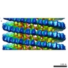 5027MC M: map data used to model this data C: citing same article ( |
|---|---|
| Similar structure data |

-Links
 PDBj
PDBj


- Assembly
| Deposited unit | 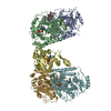
|
|---|---|
| 1 | x 33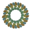
|
| 2 |
|
| 3 | 
|
| Symmetry | Helical symmetry: (Circular symmetry: 1 / Dyad axis: no / N subunits divisor: 1 / Num. of operations: 33 / Rise per n subunits: 5.506 Å / Rotation per n subunits: 168.1 °) |
-Components
-Tubulin alpha-1A ... , 2 types, 2 molecules AF
| #1: Protein | Mass: 50107.238 Da / Num. of mol.: 1 / Source method: isolated from a natural source / Source: (natural) |
|---|---|
| #4: Protein | Mass: 50163.344 Da / Num. of mol.: 1 / Source method: isolated from a natural source / Source: (natural) |
-Protein , 2 types, 3 molecules BGD
| #2: Protein | Mass: 49907.770 Da / Num. of mol.: 2 / Source method: isolated from a natural source / Source: (natural) #3: Protein | | Mass: 37072.609 Da / Num. of mol.: 1 Source method: isolated from a genetically manipulated source Source: (gene. exp.)  |
|---|
-Non-polymers , 8 types, 118 molecules 


| #5: Chemical | ChemComp-ZN /  Mass: 65.409 Da / Num. of mol.: 1 / Source method: obtained synthetically / Formula: Zn Mass: 65.409 Da / Num. of mol.: 1 / Source method: obtained synthetically / Formula: Zn | ||||||||||||
|---|---|---|---|---|---|---|---|---|---|---|---|---|---|
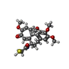
| #6: Chemical |  Mass: 24.305 Da / Num. of mol.: 3 / Source method: obtained synthetically / Formula: Mg Mass: 24.305 Da / Num. of mol.: 3 / Source method: obtained synthetically / Formula: Mg#7: Chemical |  Mass: 523.180 Da / Num. of mol.: 2 / Source method: obtained synthetically / Formula: C10H16N5O14P3 / Comment: GTP, energy-carrying molecule*YM Mass: 523.180 Da / Num. of mol.: 2 / Source method: obtained synthetically / Formula: C10H16N5O14P3 / Comment: GTP, energy-carrying molecule*YM#8: Chemical |  Type: RNA linking / Mass: 443.201 Da / Num. of mol.: 2 / Source method: obtained synthetically / Formula: C10H15N5O11P2 / Comment: GDP, energy-carrying molecule*YM Type: RNA linking / Mass: 443.201 Da / Num. of mol.: 2 / Source method: obtained synthetically / Formula: C10H15N5O11P2 / Comment: GDP, energy-carrying molecule*YM#9: Chemical | ChemComp-TA1 / |  Mass: 853.906 Da / Num. of mol.: 1 / Source method: obtained synthetically / Formula: C47H51NO14 / Comment: medication, chemotherapy*YM Mass: 853.906 Da / Num. of mol.: 1 / Source method: obtained synthetically / Formula: C47H51NO14 / Comment: medication, chemotherapy*YM#10: Chemical | ChemComp-ANP / |  Mass: 506.196 Da / Num. of mol.: 1 / Source method: obtained synthetically / Formula: C10H17N6O12P3 / Comment: AMP-PNP, energy-carrying molecule analogue*YM Mass: 506.196 Da / Num. of mol.: 1 / Source method: obtained synthetically / Formula: C10H17N6O12P3 / Comment: AMP-PNP, energy-carrying molecule analogue*YM#11: Chemical | ChemComp-CN2 / |  Mass: 431.502 Da / Num. of mol.: 1 / Source method: obtained synthetically / Formula: C22H25NO6S Mass: 431.502 Da / Num. of mol.: 1 / Source method: obtained synthetically / Formula: C22H25NO6S#12: Water | ChemComp-HOH / | Mass: 18.015 Da / Num. of mol.: 107 / Source method: isolated from a natural source / Formula: H2O |
-Details
| Sequence details | DBREF IS NOT PROVIDED BECAUSE THE STRUCTURES USED TO GENERATE THE MODELS WERE DERIVED FROM ...DBREF IS NOT PROVIDED BECAUSE THE STRUCTURES |
|---|
-Experimental details
-Experiment
| Experiment | Method: ELECTRON MICROSCOPY |
|---|---|
| EM experiment | Aggregation state: FILAMENT / 3D reconstruction method: helical reconstruction |
- Sample preparation
| Component | Name: Kinesin13-Microtubule ring complex / Type: COMPLEX |
|---|---|
| Buffer solution | pH: 6.8 Details: 80 mM Pipes, pH 6.8, 2 mM MgCl2, 1 mM EGTA, 2 mM AMP-PNP, 0.02 mM taxol |
| Specimen | Embedding applied: NO / Shadowing applied: NO / Staining applied: NO / Vitrification applied: YES |
| Specimen support | Details: 400 mesh Quantifoil grid R2/4 |
| Vitrification | Instrument: FEI VITROBOT MARK I / Cryogen name: ETHANE |
- Electron microscopy imaging
| Experimental equipment |  Model: Tecnai F20 / Image courtesy: FEI Company |
|---|---|
| Microscopy | Model: FEI TECNAI F20 |
| Electron gun | Electron source:  FIELD EMISSION GUN / Accelerating voltage: 200 kV / Illumination mode: FLOOD BEAM FIELD EMISSION GUN / Accelerating voltage: 200 kV / Illumination mode: FLOOD BEAM |
| Electron lens | Mode: BRIGHT FIELD / Nominal magnification: 50000 X / Nominal defocus max: 2500 nm / Nominal defocus min: 1500 nm |
| Image recording | Film or detector model: KODAK SO-163 FILM |
- Processing
| EM software |
| ||||||||||||||||||||||||
|---|---|---|---|---|---|---|---|---|---|---|---|---|---|---|---|---|---|---|---|---|---|---|---|---|---|
| CTF correction | Details: Each particle | ||||||||||||||||||||||||
| 3D reconstruction | Method: HELICAL / Resolution: 28 Å / Nominal pixel size: 2.5 Å / Details: Helical Fourier-Bessel / Symmetry type: HELICAL | ||||||||||||||||||||||||
| Atomic model building | Space: REAL | ||||||||||||||||||||||||
| Refinement step | Cycle: LAST
|
