Movie
Movie Controller


+ Open data

- Basic information
| Entry | Database: EMDB / ID: EMD-5027 | |||||||||
|---|---|---|---|---|---|---|---|---|---|---|
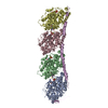
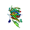
| Title | Kinesin13-Microtubule Ring Complex | |||||||||
 Map data Map data | Kinesin 13-Microtubule Ring Complex | |||||||||
 Sample Sample |
| |||||||||
 Keywords Keywords | Kinesin / Kinesin-13 / microtubule / tubulin / depolymerization / Kin-I / M-Kinesin / cytoskeleton | |||||||||
| Function / homology |  Function and homology information Function and homology informationpostsynaptic cytoskeleton organization / regulation of chromosome segregation / establishment or maintenance of microtubule cytoskeleton polarity / Amplification of signal from unattached kinetochores via a MAD2 inhibitory signal / Kinesins / metaphase chromosome alignment / Mitotic Prometaphase / EML4 and NUDC in mitotic spindle formation / Resolution of Sister Chromatid Cohesion / COPI-dependent Golgi-to-ER retrograde traffic ...postsynaptic cytoskeleton organization / regulation of chromosome segregation / establishment or maintenance of microtubule cytoskeleton polarity / Amplification of signal from unattached kinetochores via a MAD2 inhibitory signal / Kinesins / metaphase chromosome alignment / Mitotic Prometaphase / EML4 and NUDC in mitotic spindle formation / Resolution of Sister Chromatid Cohesion / COPI-dependent Golgi-to-ER retrograde traffic / RHO GTPases Activate Formins / Separation of Sister Chromatids / microtubule plus-end / attachment of mitotic spindle microtubules to kinetochore / MHC class II antigen presentation / microtubule plus-end binding / microtubule depolymerization / microtubule motor activity / kinesin complex / microtubule-based movement / mitotic metaphase chromosome alignment / tubulin complex / regulation of neurotransmitter receptor localization to postsynaptic specialization membrane / chromosome, centromeric region / structural constituent of cytoskeleton / kinetochore / microtubule cytoskeleton organization / spindle / neuron migration / mitotic cell cycle / presynapse / microtubule cytoskeleton / microtubule binding / microtubule / Hydrolases; Acting on acid anhydrides; Acting on GTP to facilitate cellular and subcellular movement / postsynapse / cell division / hydrolase activity / GTPase activity / centrosome / GTP binding / glutamatergic synapse / ATP hydrolysis activity / ATP binding / metal ion binding / nucleus / cytoplasm Similarity search - Function | |||||||||
| Method | helical reconstruction / cryo EM / Resolution: 28.0 Å | |||||||||
 Authors Authors | Tan D / Rice WJ / Sosa H | |||||||||
 Citation Citation | Journal: Structure / Year: 2008 Title: Structure of the kinesin13-microtubule ring complex. Authors: Dongyan Tan / William J Rice / Hernando Sosa /  Abstract: To investigate the mechanism of kinesin13-induced microtubule depolymerization, we have calculated a three-dimensional (3D) map of the kinesin13-microtubule ring complex, using cryo-electron ...To investigate the mechanism of kinesin13-induced microtubule depolymerization, we have calculated a three-dimensional (3D) map of the kinesin13-microtubule ring complex, using cryo-electron microscopy (cryo-EM) and image analysis. An atomic model of the complex was produced by docking the crystal structures of tubulin and a kinesin13 motor domain (MD) into the 3D map. The model reveals a snapshot of the depolymerization mechanism by providing a 3D view of the complex formed between the kinesin13 MD and a curved tubulin protofilament (pf). It suggests that contacts mediated by kinesin13 class-specific residues in the putative microtubule-binding site stabilize intra-dimer tubulin curvature. In addition, a tubulin-binding site on the kinesin13 MD was identified. Mutations at this class-conserved site selectively disrupt the formation of microtubule-associated ring complexes. | |||||||||
| History |
|
- Structure visualization
| Movie |
Movie viewer |
|---|---|
| Structure viewer | EM map: SurfViewMolmilJmol/JSmol |
| Supplemental images |

- Downloads & links
-EMDB archive
| Map data | emd_5027.map.gz | 340.9 KB | EMDB map data format | |
|---|---|---|---|---|
| Header (meta data) | emd-5027-v30.xmlemd-5027.xml | 14.3 KB 14.3 KB | Display Display | EMDB header |
| Images | emd_5027_1.tif | 747.2 KB | ||
| Archive directory |  http://ftp.pdbj.org/pub/emdb/structures/EMD-5027ftp://ftp.pdbj.org/pub/emdb/structures/EMD-5027 http://ftp.pdbj.org/pub/emdb/structures/EMD-5027ftp://ftp.pdbj.org/pub/emdb/structures/EMD-5027 | HTTPS FTP |
-Related structure data
| Related structure data |  3edlMC M: atomic model generated by this map C: citing same article ( |
|---|---|
| Similar structure data |


-Links
| EMDB pages | EMDB (EBI/PDBe) / EMDataResource |
|---|---|
| Related items in Molecule of the Month |


-Map
| File | Download / File: emd_5027.map.gz / Format: CCP4 / Size: 1.6 MB / Type: IMAGE STORED AS FLOATING POINT NUMBER (4 BYTES) | ||||||||||||||||||||||||||||||||||||||||||||||||||||||||||||||||||||
|---|---|---|---|---|---|---|---|---|---|---|---|---|---|---|---|---|---|---|---|---|---|---|---|---|---|---|---|---|---|---|---|---|---|---|---|---|---|---|---|---|---|---|---|---|---|---|---|---|---|---|---|---|---|---|---|---|---|---|---|---|---|---|---|---|---|---|---|---|---|
| Annotation | Kinesin 13-Microtubule Ring Complex | ||||||||||||||||||||||||||||||||||||||||||||||||||||||||||||||||||||
| Projections & slices | Image control
Images are generated by Spider. generated in cubic-lattice coordinate | ||||||||||||||||||||||||||||||||||||||||||||||||||||||||||||||||||||
| Voxel size | X=Y=Z: 6 Å | ||||||||||||||||||||||||||||||||||||||||||||||||||||||||||||||||||||
| Density |
| ||||||||||||||||||||||||||||||||||||||||||||||||||||||||||||||||||||
| Symmetry | Space group: 1 | ||||||||||||||||||||||||||||||||||||||||||||||||||||||||||||||||||||
| Details | EMDB XML:
CCP4 map header:
| ||||||||||||||||||||||||||||||||||||||||||||||||||||||||||||||||||||
 Z (Sec.)
 Y (Row.)
 X (Col.)


-Supplemental data
- Sample components
-Entire : Kinesin13-Microtubule Ring Complex
| Entire | Name: Kinesin13-Microtubule Ring Complex |
|---|---|
| Components |
|
-Supramolecule #1000: Kinesin13-Microtubule Ring Complex
| Supramolecule | Name: Kinesin13-Microtubule Ring Complex / type: sample / ID: 1000 / Number unique components: 3 |
|---|---|
| Molecular weight | Experimental: 250 KDa / Theoretical: 250 KDa / Method: SDS-PAGE |
-Macromolecule #1: KLP10a
| Macromolecule | Name: KLP10a / type: protein_or_peptide / ID: 1 / Name.synonym: Kinesin-13 / Number of copies: 1 / Oligomeric state: monomer / Recombinant expression: Yes |
|---|---|
| Source (natural) | Cell: BL21 / Location in cell: cytosol |
| Molecular weight | Experimental: 400 KDa / Theoretical: 400 KDa |
| Recombinant expression | Organism:  |
-Macromolecule #2: alpha-tubulin
| Macromolecule | Name: alpha-tubulin / type: protein_or_peptide / ID: 2 / Name.synonym: tubulin / Number of copies: 2 / Oligomeric state: Heterodimer / Recombinant expression: No / Database: NCBI |
|---|---|
| Source (natural) | Tissue: Bos taurus Brain |
| Molecular weight | Experimental: 550 KDa |
| Sequence | GO: tubulin complex / InterPro: Alpha tubulin |
-Macromolecule #3: beta-tubulin
| Macromolecule | Name: beta-tubulin / type: protein_or_peptide / ID: 3 / Name.synonym: tubulin / Number of copies: 2 / Oligomeric state: Hterodimer / Recombinant expression: No / Database: NCBI |
|---|---|
| Source (natural) | Tissue: Brain |
| Molecular weight | Experimental: 550 KDa |
| Sequence | GO: tubulin complex / InterPro: Beta tubulin |
-Experimental details
-Structure determination
| Method | cryo EM |
|---|---|
 Processing Processing | helical reconstruction |
| Aggregation state | filament |
-Sample preparation
| Concentration | 0.6 mg/mL |
|---|---|
| Buffer | pH: 6.8 Details: 80 mM Pipes, pH 6.8, 2 mM MgCl2, 1 mM EGTA, 2 mM AMP-PNP, 0.02 mM taxol |
| Grid | Details: Quantifoil grids |
| Vitrification | Cryogen name: ETHANE / Chamber humidity: 100 % / Instrument: OTHER / Details: Vitrification instrument: VitroBot Method: Blot for 2 seconds, Blot Offset (mm) -2, Plunge Time 1 sec, Wait Time 0 sec |
- Electron microscopy #1
| Microscopy ID | 1 |
|---|---|
| Microscope | FEI TECNAI 20 |
| Image recording | Category: FILM / Film or detector model: KODAK SO-163 FILM / Digitization - Scanner: OTHER / Digitization - Sampling interval: 12.5 µm / Number real images: 5 / Average electron dose: 9 e/Å2 / Bits/pixel: 16 |
| Electron beam | Acceleration voltage: 120 kV / Electron source: LAB6 |
| Electron optics | Illumination mode: FLOOD BEAM / Imaging mode: BRIGHT FIELD / Nominal defocus max: 2.5 µm / Nominal defocus min: 1.5 µm / Nominal magnification: 50000 |
| Sample stage | Specimen holder: Side entry liquid nitrogen-cooled cryo specimen holder Specimen holder model: GATAN LIQUID NITROGEN |
-Electron microscopy #2
| Microscopy ID | 2 |
|---|---|
| Microscope | FEI TECNAI F20 |
| Image recording | Category: FILM / Film or detector model: KODAK SO-163 FILM / Digitization - Scanner: OTHER / Digitization - Sampling interval: 12.5 µm / Number real images: 5 / Average electron dose: 9 e/Å2 / Bits/pixel: 16 |
| Electron beam | Acceleration voltage: 200 kV / Electron source:  FIELD EMISSION GUN FIELD EMISSION GUN |
| Electron optics | Illumination mode: FLOOD BEAM / Imaging mode: BRIGHT FIELD / Nominal magnification: 50000 |
| Sample stage | Specimen holder: Side entry liquid nitrogen-cooled cryo specimen holder Specimen holder model: GATAN LIQUID NITROGEN |
| Experimental equipment |  Model: Tecnai F20 / Image courtesy: FEI Company |
-Image processing
| Final reconstruction | Algorithm: OTHER / Resolution.type: BY AUTHOR / Resolution: 28.0 Å / Resolution method: FSC 0.5 CUT-OFF / Software - Name: MRC, SUPRIM, Phoelix, Other Details: Final map is the averaged near and far layer line data from 5 filaments (10 datasets, 3296 asymmetric units) |
|---|---|
| CTF correction | Details: Each Particle |
-Atomic model buiding 1
| Initial model | PDB ID: Chain - Chain ID: A |
|---|---|
| Software | Name: COLORES |
| Details | PDBEntryID_givenInChain. Protocol: Rigid-Body |
| Refinement | Space: REAL / Protocol: RIGID BODY FIT |
| Output model | PDB-3edl: |
-Atomic model buiding 2
| Initial model | PDB ID: |
|---|---|
| Software | Name: COLORES |
| Details | Protocol: Rigid Body |
| Refinement | Space: REAL / Protocol: RIGID BODY FIT |
| Output model | PDB-3edl: |