 Movie
Movie Controller

[English] 日本語
 Yorodumi
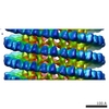
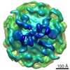
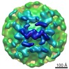
- PDB-3j2u: Kinesin-13 KLP10A HD in complex with CS-tubulin and a microtubule -
+ Open data

- Basic information
| Entry | Database: PDB / ID: 3j2u | ||||||
|---|---|---|---|---|---|---|---|
| Title | Kinesin-13 KLP10A HD in complex with CS-tubulin and a microtubule | ||||||
 Components Components |
| ||||||
 Keywords Keywords | MOTOR PROTEIN / tubulin / kinesin / kinesin-13 / KinI / depolymerase / depolymerization / microtubule / Kinesin13 | ||||||
| Function / homology |  Function and homology information Function and homology informationestablishment of mitotic spindle asymmetry / establishment of meiotic spindle orientation / plus-end specific microtubule depolymerization / cortical microtubule / asymmetric protein localization involved in cell fate determination / meiotic spindle pole / mitotic spindle astral microtubule / COPI-dependent Golgi-to-ER retrograde traffic / Kinesins / centriole assembly ...establishment of mitotic spindle asymmetry / establishment of meiotic spindle orientation / plus-end specific microtubule depolymerization / cortical microtubule / asymmetric protein localization involved in cell fate determination / meiotic spindle pole / mitotic spindle astral microtubule / COPI-dependent Golgi-to-ER retrograde traffic / Kinesins / centriole assembly / kinetochore microtubule / meiotic spindle organization / spindle assembly involved in female meiosis I / non-motile cilium assembly / plus-end-directed microtubule motor activity / positive regulation of axon guidance / meiotic spindle / microtubule depolymerization / spindle organization / microtubule motor activity / kinesin complex / microtubule-based movement / mitotic spindle pole / centrosome duplication / cytoskeletal motor activity / chromosome, centromeric region / microtubule-based process / cytoplasmic microtubule / cellular response to interleukin-4 / mitotic spindle organization / microtubule cytoskeleton organization / structural constituent of cytoskeleton / neuron migration / spindle / spindle pole / mitotic cell cycle / double-stranded RNA binding / microtubule cytoskeleton / microtubule binding / microtubule / Hydrolases; Acting on acid anhydrides; Acting on GTP to facilitate cellular and subcellular movement / cilium / protein heterodimerization activity / cell division / GTPase activity / centrosome / ubiquitin protein ligase binding / GTP binding / ATP hydrolysis activity / ATP binding / metal ion binding / cytosol / cytoplasm Similarity search - Function | ||||||
| Biological species |   | ||||||
| Method | ELECTRON MICROSCOPY / helical reconstruction / cryo EM / Resolution: 10.8 Å | ||||||
 Authors Authors | Asenjo, A.B. / Chatterjee, C. / Tan, D. / DePaoli, V. / Rice, W.J. / Diaz-Avalos, R. / Silvestry, M. / Sosa, H. | ||||||
 Citation Citation | Journal: Cell Rep / Year: 2013 Title: Structural model for tubulin recognition and deformation by kinesin-13 microtubule depolymerases. Authors: Ana B Asenjo / Chandrima Chatterjee / Dongyan Tan / Vania DePaoli / William J Rice / Ruben Diaz-Avalos / Mariena Silvestry / Hernando Sosa /  Abstract: To elucidate the structural basis of the mechanism of microtubule depolymerization by kinesin-13s, we analyzed complexes of tubulin and the Drosophila melanogaster kinesin-13 KLP10A by electron ...To elucidate the structural basis of the mechanism of microtubule depolymerization by kinesin-13s, we analyzed complexes of tubulin and the Drosophila melanogaster kinesin-13 KLP10A by electron microscopy (EM) and fluorescence polarization microscopy. We report a nanometer-resolution (1.1 nm) cryo-EM three-dimensional structure of the KLP10A head domain (KLP10AHD) bound to curved tubulin. We found that binding of KLP10AHD induces a distinct tubulin configuration with displacement (shear) between tubulin subunits in addition to curvature. In this configuration, the kinesin-binding site differs from that in straight tubulin, providing an explanation for the distinct interaction modes of kinesin-13s with the microtubule lattice or its ends. The KLP10AHD-tubulin interface comprises three areas of interaction, suggesting a crossbow-type tubulin-bending mechanism. These areas include the kinesin-13 family conserved KVD residues, and as predicted from the crossbow model, mutating these residues changes the orientation and mobility of KLP10AHDs interacting with the microtubule. | ||||||
| History |
|
- Structure visualization
| Movie |
Movie viewer |
|---|---|
| Structure viewer | Molecule: MolmilJmol/JSmol |
 UCSF Chimera
- Downloads & links
-Download
| PDBx/mmCIF format | 3j2u.cif.gz | 393 KB | Display | PDBx/mmCIF format |
|---|---|---|---|---|
| PDB format | pdb3j2u.ent.gz | 312.8 KB | Display | PDB format |
| PDBx/mmJSON format | 3j2u.json.gz | Tree view | PDBx/mmJSON format | |
| Others |  Other downloads Other downloads |
-Validation report
| Arichive directory | https://data.pdbj.org/pub/pdb/validation_reports/j2/3j2uftp://data.pdbj.org/pub/pdb/validation_reports/j2/3j2u | HTTPS FTP |
|---|
-Related structure data
| Related structure data | 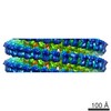 5565MC M: map data used to model this data C: citing same article ( |
|---|---|
| Similar structure data |
-Links
 PDBj
PDBj


- Assembly
| Deposited unit | 
|
|---|---|
| 1 | x 42
|
| 2 |
|
| 3 | 
|
| Symmetry | Helical symmetry: (Circular symmetry: 1 / Dyad axis: no / N subunits divisor: 1 / Num. of operations: 42 / Rise per n subunits: 5.553 Å / Rotation per n subunits: 168.087 °) |
-Components
| #1: Protein | Mass: 41810.918 Da / Num. of mol.: 1 / Fragment: head domain (UNP residues 279-615) Source method: isolated from a genetically manipulated source Source: (gene. exp.)  | ||
|---|---|---|---|
| #2: Protein | Mass: 50107.238 Da / Num. of mol.: 2 / Source method: isolated from a natural source / Source: (natural) #3: Protein | Mass: 49907.770 Da / Num. of mol.: 2 / Source method: isolated from a natural source / Source: (natural) |
-Experimental details
-Experiment
| Experiment | Method: ELECTRON MICROSCOPY |
|---|---|
| EM experiment | Aggregation state: FILAMENT / 3D reconstruction method: helical reconstruction |
- Sample preparation
| Component | Name: kinesin-13 KLP10A head domain in complex with CS-tubulin and a microtubule Type: COMPLEX |
|---|---|
| Specimen | Embedding applied: NO / Shadowing applied: NO / Staining applied: NO / Vitrification applied: YES |
| Vitrification | Instrument: FEI VITROBOT MARK I / Cryogen name: ETHANE |
- Electron microscopy imaging
| Experimental equipment |  Model: Tecnai F20 / Image courtesy: FEI Company | |||||||||||||||
|---|---|---|---|---|---|---|---|---|---|---|---|---|---|---|---|---|
| EM imaging | Date: May 1, 2012 / Electron source:
|
- Processing
| EM software |
| ||||||||||||||||||||||||
|---|---|---|---|---|---|---|---|---|---|---|---|---|---|---|---|---|---|---|---|---|---|---|---|---|---|
| CTF correction | Details: each particle | ||||||||||||||||||||||||
| Helical symmerty | Angular rotation/subunit: 168.087 ° / Axial rise/subunit: 5.553 Å / Axial symmetry: C1 | ||||||||||||||||||||||||
| 3D reconstruction | Resolution: 10.8 Å / Resolution method: FSC 0.143 CUT-OFF / Num. of particles: 54584 / Nominal pixel size: 2 Å / Actual pixel size: 2 Å / Refinement type: HALF-MAPS REFINED INDEPENDENTLY / Symmetry type: HELICAL | ||||||||||||||||||||||||
| Atomic model building | Protocol: RIGID BODY FIT / Space: REAL Details: REFINEMENT PROTOCOL--RIGID BODY DETAILS--The domains were separately fitted with the global fitting option of the routine fitmap within UCSF-Chimera. Best fit was chosen based on the maximum ...Details: REFINEMENT PROTOCOL--RIGID BODY DETAILS--The domains were separately fitted with the global fitting option of the routine fitmap within UCSF-Chimera. Best fit was chosen based on the maximum cross-correlation value between the EM density and an 11 A resolution density model of the atomic structure of the domain to be fitted. | ||||||||||||||||||||||||
| Atomic model building | 3D fitting-ID: 1 / Accession code: 1JFF / Initial refinement model-ID: 1 / PDB-ID: 1JFF / Source name: PDB / Type: experimental model
| ||||||||||||||||||||||||
| Refinement step | Cycle: LAST
|

