 Movie
Movie Controller


+ Open data

- Basic information
| Entry | Database: PDB / ID: 6b0c | ||||||
|---|---|---|---|---|---|---|---|
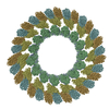
| Title | KLP10A-AMPPNP in complex with curved tubulin and a microtubule | ||||||
 Components Components |
| ||||||
 Keywords Keywords | MOTOR PROTEIN/STRUCTURAL PROTEIN / kinesin 13 / microtubule / tubulin / depolymerization / MOTOR PROTEIN-STRUCTURAL PROTEIN complex | ||||||
| Function / homology |  Function and homology information Function and homology informationestablishment of mitotic spindle asymmetry / establishment of meiotic spindle orientation / plus-end specific microtubule depolymerization / cortical microtubule / asymmetric protein localization involved in cell fate determination / COPI-dependent Golgi-to-ER retrograde traffic / Kinesins / meiotic spindle pole / mitotic spindle astral microtubule / centriole assembly ...establishment of mitotic spindle asymmetry / establishment of meiotic spindle orientation / plus-end specific microtubule depolymerization / cortical microtubule / asymmetric protein localization involved in cell fate determination / COPI-dependent Golgi-to-ER retrograde traffic / Kinesins / meiotic spindle pole / mitotic spindle astral microtubule / centriole assembly / kinetochore microtubule / meiotic spindle organization / spindle assembly involved in female meiosis I / non-motile cilium assembly / plus-end-directed microtubule motor activity / Microtubule-dependent trafficking of connexons from Golgi to the plasma membrane / Resolution of Sister Chromatid Cohesion / Hedgehog 'off' state / Cilium Assembly / Intraflagellar transport / COPI-dependent Golgi-to-ER retrograde traffic / Mitotic Prometaphase / Carboxyterminal post-translational modifications of tubulin / RHOH GTPase cycle / EML4 and NUDC in mitotic spindle formation / Sealing of the nuclear envelope (NE) by ESCRT-III / Kinesins / PKR-mediated signaling / Separation of Sister Chromatids / The role of GTSE1 in G2/M progression after G2 checkpoint / Aggrephagy / meiotic spindle / microtubule depolymerization / RHO GTPases activate IQGAPs / RHO GTPases Activate Formins / HSP90 chaperone cycle for steroid hormone receptors (SHR) in the presence of ligand / MHC class II antigen presentation / spindle organization / Recruitment of NuMA to mitotic centrosomes / microtubule motor activity / kinesin complex / COPI-mediated anterograde transport / microtubule-based movement / mitotic spindle pole / cytoskeletal motor activity / centrosome duplication / chromosome, centromeric region / microtubule-based process / mitotic spindle organization / neuron migration / microtubule cytoskeleton organization / spindle / structural constituent of cytoskeleton / spindle pole / microtubule cytoskeleton / mitotic cell cycle / microtubule binding / microtubule / Hydrolases; Acting on acid anhydrides; Acting on GTP to facilitate cellular and subcellular movement / cell division / GTPase activity / centrosome / GTP binding / ATP hydrolysis activity / ATP binding / metal ion binding / cytoplasm Similarity search - Function | ||||||
| Biological species |   | ||||||
| Method | ELECTRON MICROSCOPY / helical reconstruction / cryo EM / Resolution: 3.51 Å | ||||||
 Authors Authors | Benoit, M.P.M.H. / Asenjo, A.B. / Sosa, H. | ||||||
| Funding support |  United States, 1items United States, 1items
| ||||||
 Citation Citation | Journal: Nat Commun / Year: 2018 Title: Cryo-EM reveals the structural basis of microtubule depolymerization by kinesin-13s. Authors: Matthieu P M H Benoit / Ana B Asenjo / Hernando Sosa / Abstract: Kinesin-13s constitute a distinct group within the kinesin superfamily of motor proteins that promote microtubule depolymerization and lack motile activity. The molecular mechanism by which kinesin- ...Kinesin-13s constitute a distinct group within the kinesin superfamily of motor proteins that promote microtubule depolymerization and lack motile activity. The molecular mechanism by which kinesin-13s depolymerize microtubules and are adapted to perform a seemingly very different activity from other kinesins is still unclear. To address this issue, here we report the near atomic resolution cryo-electron microscopy (cryo-EM) structures of Drosophila melanogaster kinesin-13 KLP10A protein constructs bound to curved or straight tubulin in different nucleotide states. These structures show how nucleotide induced conformational changes near the catalytic site are coupled with movement of the kinesin-13-specific loop-2 to induce tubulin curvature leading to microtubule depolymerization. The data highlight a modular structure that allows similar kinesin core motor-domains to be used for different functions, such as motility or microtubule depolymerization. | ||||||
| History |
|
- Structure visualization
| Movie |
Movie viewer |
|---|---|
| Structure viewer | Molecule: MolmilJmol/JSmol |
- Downloads & links
-Download
| PDBx/mmCIF format | 6b0c.cif.gz | 374.2 KB | Display | PDBx/mmCIF format |
|---|---|---|---|---|
| PDB format | pdb6b0c.ent.gz | 301.7 KB | Display | PDB format |
| PDBx/mmJSON format | 6b0c.json.gz | Tree view | PDBx/mmJSON format | |
| Others |  Other downloads Other downloads |
-Validation report
| Arichive directory | https://data.pdbj.org/pub/pdb/validation_reports/b0/6b0cftp://data.pdbj.org/pub/pdb/validation_reports/b0/6b0c | HTTPS FTP |
|---|
-Related structure data
| Related structure data |  7026MC 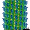 7027C 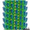 7028C 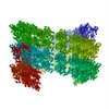 6b0iC  6b0lC M: map data used to model this data C: citing same article ( |
|---|---|
| Similar structure data |
-Links
 PDBj
PDBj


- Assembly
| Deposited unit | 
|
|---|---|
| 1 | x 35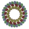
|
| 2 |
|
| 3 | 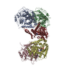
|
| Symmetry | Helical symmetry: (Circular symmetry: 1 / Dyad axis: no / N subunits divisor: 1 / Num. of operations: 35 / Rise per n subunits: 5.57 Å / Rotation per n subunits: 168.089 °) |
-Components
-Protein , 3 types, 5 molecules ACBDK
| #1: Protein | Mass: 50204.445 Da / Num. of mol.: 2 / Source method: isolated from a natural source / Source: (natural) #2: Protein | Mass: 49999.887 Da / Num. of mol.: 2 / Source method: isolated from a natural source / Source: (natural) #3: Protein | | Mass: 41810.918 Da / Num. of mol.: 1 / Fragment: motor (UNP residues 279-615) Source method: isolated from a genetically manipulated source Source: (gene. exp.)  |
|---|
-Non-polymers , 5 types, 9 molecules 


| #4: Chemical |  Mass: 523.180 Da / Num. of mol.: 2 / Source method: obtained synthetically / Formula: C10H16N5O14P3 / Comment: GTP, energy-carrying molecule*YM Mass: 523.180 Da / Num. of mol.: 2 / Source method: obtained synthetically / Formula: C10H16N5O14P3 / Comment: GTP, energy-carrying molecule*YM#5: Chemical |  Mass: 24.305 Da / Num. of mol.: 3 Mass: 24.305 Da / Num. of mol.: 3Source method: isolated from a genetically manipulated source Formula: Mg #6: Chemical |  Type: RNA linking / Mass: 443.201 Da / Num. of mol.: 2 / Source method: obtained synthetically / Formula: C10H15N5O11P2 / Comment: GDP, energy-carrying molecule*YM Type: RNA linking / Mass: 443.201 Da / Num. of mol.: 2 / Source method: obtained synthetically / Formula: C10H15N5O11P2 / Comment: GDP, energy-carrying molecule*YM#7: Chemical | ChemComp-TA1 / |  Mass: 853.906 Da / Num. of mol.: 1 / Source method: obtained synthetically / Formula: C47H51NO14 / Comment: medication, chemotherapy*YM Mass: 853.906 Da / Num. of mol.: 1 / Source method: obtained synthetically / Formula: C47H51NO14 / Comment: medication, chemotherapy*YM#8: Chemical | ChemComp-ANP / |  Mass: 506.196 Da / Num. of mol.: 1 / Source method: obtained synthetically / Formula: C10H17N6O12P3 / Comment: AMP-PNP, energy-carrying molecule analogue*YM Mass: 506.196 Da / Num. of mol.: 1 / Source method: obtained synthetically / Formula: C10H17N6O12P3 / Comment: AMP-PNP, energy-carrying molecule analogue*YM |
|---|
-Experimental details
-Experiment
| Experiment | Method: ELECTRON MICROSCOPY |
|---|---|
| EM experiment | Aggregation state: FILAMENT / 3D reconstruction method: helical reconstruction |
- Sample preparation
| Component |
| ||||||||||||||||||||||||||||
|---|---|---|---|---|---|---|---|---|---|---|---|---|---|---|---|---|---|---|---|---|---|---|---|---|---|---|---|---|---|
| Molecular weight | Experimental value: NO | ||||||||||||||||||||||||||||
| Source (natural) |
| ||||||||||||||||||||||||||||
| Source (recombinant) | Organism: | ||||||||||||||||||||||||||||
| Buffer solution | pH: 6.8 | ||||||||||||||||||||||||||||
| Buffer component |
| ||||||||||||||||||||||||||||
| Specimen | Embedding applied: NO / Shadowing applied: NO / Staining applied: NO / Vitrification applied: YES | ||||||||||||||||||||||||||||
| Specimen support | Grid material: COPPER | ||||||||||||||||||||||||||||
| Vitrification | Cryogen name: ETHANE |
- Electron microscopy imaging
| Experimental equipment |  Model: Titan Krios / Image courtesy: FEI Company |
|---|---|
| Microscopy | Model: FEI TITAN KRIOS |
| Electron gun | Electron source:  FIELD EMISSION GUN / Accelerating voltage: 300 kV / Illumination mode: FLOOD BEAM FIELD EMISSION GUN / Accelerating voltage: 300 kV / Illumination mode: FLOOD BEAM |
| Electron lens | Mode: BRIGHT FIELD / Nominal magnification: 22500 X / Calibrated magnification: 46598 X / Alignment procedure: COMA FREE |
| Specimen holder | Cryogen: NITROGEN / Specimen holder model: FEI TITAN KRIOS AUTOGRID HOLDER |
| Image recording | Electron dose: 1.25 e/Å2 / Detector mode: COUNTING / Film or detector model: GATAN K2 SUMMIT (4k x 4k) |
| Image scans | Movie frames/image: 50 / Used frames/image: 1-50 |
- Processing
| EM software |
| |||||||||||||||||||||||||||||||||||||||||||||||||||||||
|---|---|---|---|---|---|---|---|---|---|---|---|---|---|---|---|---|---|---|---|---|---|---|---|---|---|---|---|---|---|---|---|---|---|---|---|---|---|---|---|---|---|---|---|---|---|---|---|---|---|---|---|---|---|---|---|---|
| CTF correction | Type: PHASE FLIPPING AND AMPLITUDE CORRECTION | |||||||||||||||||||||||||||||||||||||||||||||||||||||||
| Helical symmerty | Angular rotation/subunit: 168.089 ° / Axial rise/subunit: 5.57 Å / Axial symmetry: C1 | |||||||||||||||||||||||||||||||||||||||||||||||||||||||
| Particle selection | Details: manual picking of filaments | |||||||||||||||||||||||||||||||||||||||||||||||||||||||
| 3D reconstruction | Resolution: 3.51 Å / Resolution method: FSC 0.143 CUT-OFF / Num. of particles: 16942 Details: 2 independent reconstructions, each using a distinct half of every filament. Symmetry type: HELICAL | |||||||||||||||||||||||||||||||||||||||||||||||||||||||
| Atomic model building | Protocol: FLEXIBLE FIT / Space: REAL |
