National Institutes of Health/National Institute of General Medical Sciences (NIH/NIGMS)
R01GM113164
United States
Citation
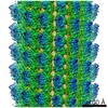
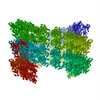
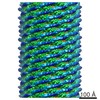
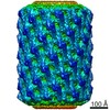
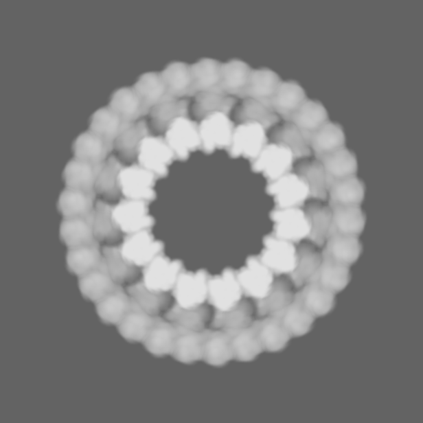
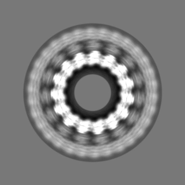
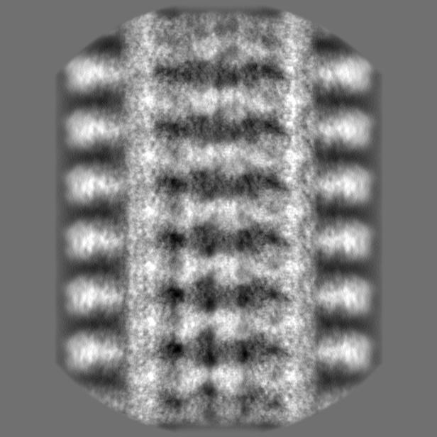
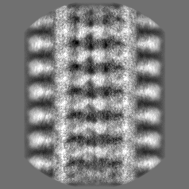
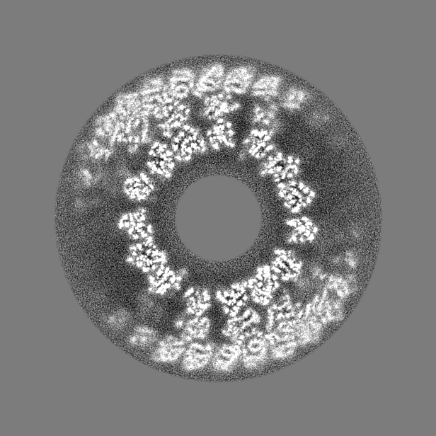
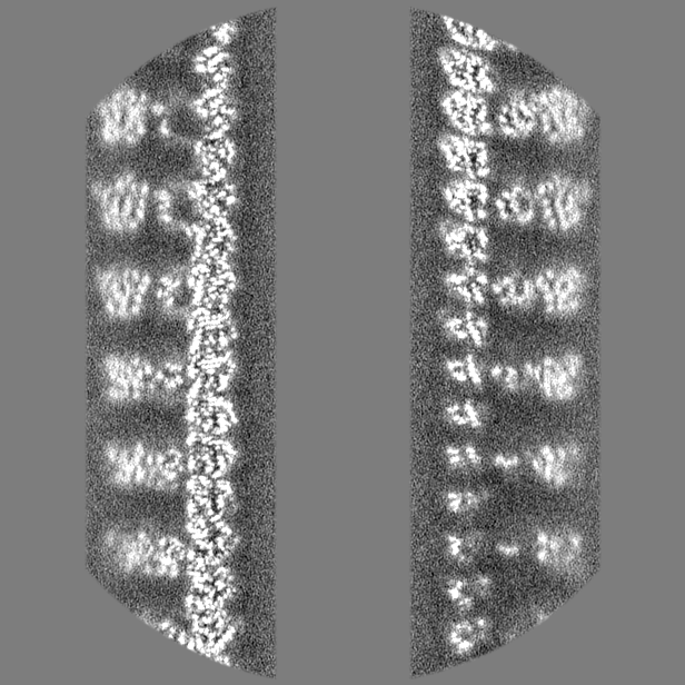
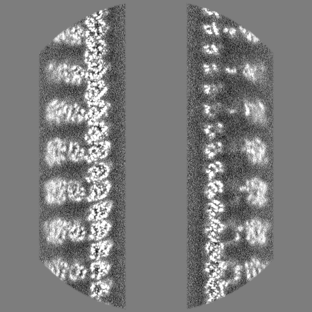
Journal: Nat Commun / Year: 2018 Title: Cryo-EM reveals the structural basis of microtubule depolymerization by kinesin-13s. Authors: Matthieu P M H Benoit / Ana B Asenjo / Hernando Sosa / Abstract: Kinesin-13s constitute a distinct group within the kinesin superfamily of motor proteins that promote microtubule depolymerization and lack motile activity. The molecular mechanism by which kinesin- ...Kinesin-13s constitute a distinct group within the kinesin superfamily of motor proteins that promote microtubule depolymerization and lack motile activity. The molecular mechanism by which kinesin-13s depolymerize microtubules and are adapted to perform a seemingly very different activity from other kinesins is still unclear. To address this issue, here we report the near atomic resolution cryo-electron microscopy (cryo-EM) structures of Drosophila melanogaster kinesin-13 KLP10A protein constructs bound to curved or straight tubulin in different nucleotide states. These structures show how nucleotide induced conformational changes near the catalytic site are coupled with movement of the kinesin-13-specific loop-2 to induce tubulin curvature leading to microtubule depolymerization. The data highlight a modular structure that allows similar kinesin core motor-domains to be used for different functions, such as motility or microtubule depolymerization.
History
Deposition
Sep 14, 2017
-
Header (metadata) release
Nov 1, 2017
-
Map release
May 2, 2018
-
Update
Mar 13, 2024
-
Current status
Mar 13, 2024
Processing site: RCSB / Status: Released
-
Structure visualization
Movie
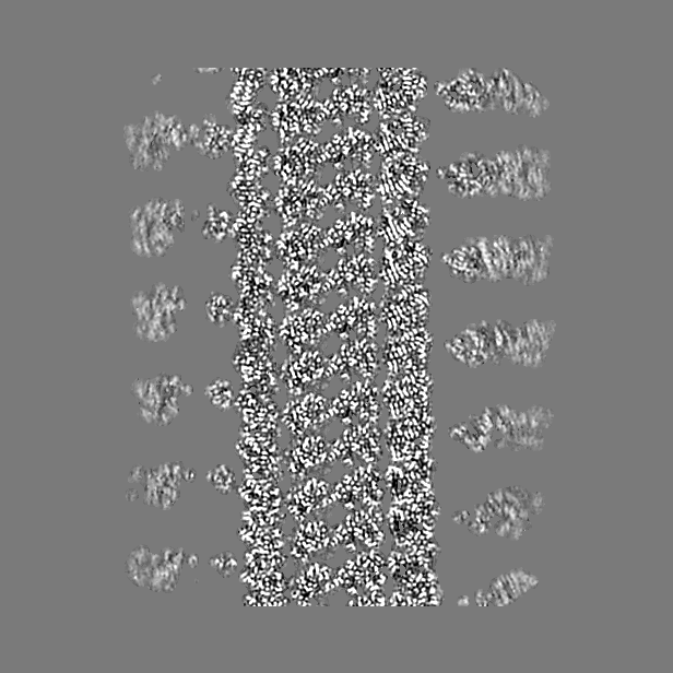
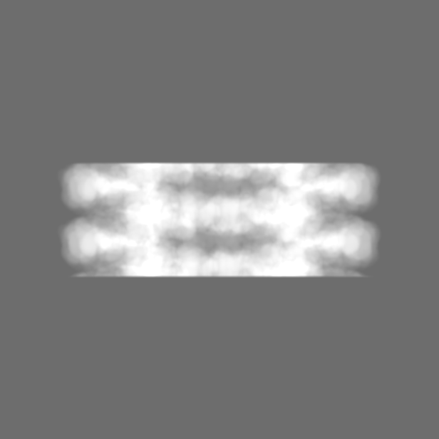
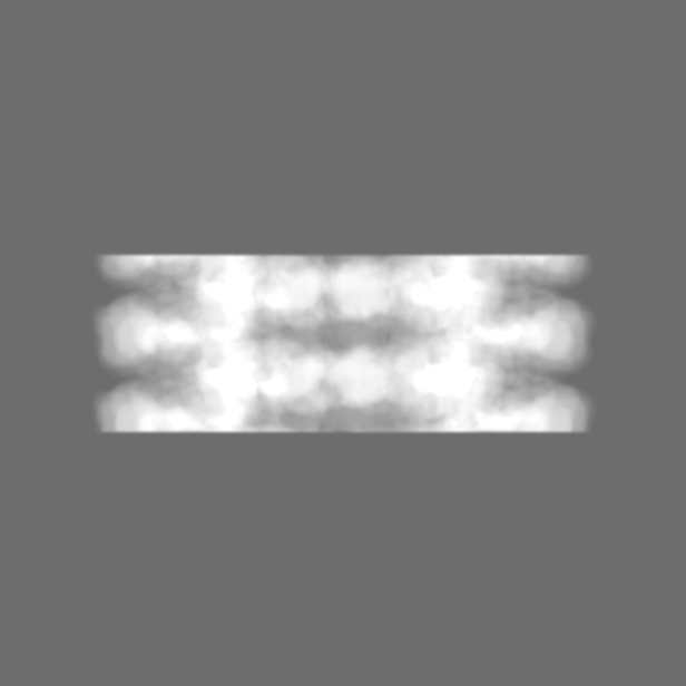
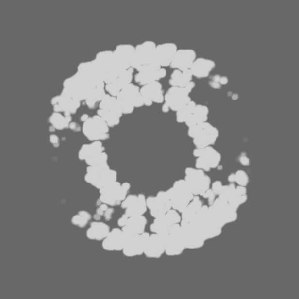
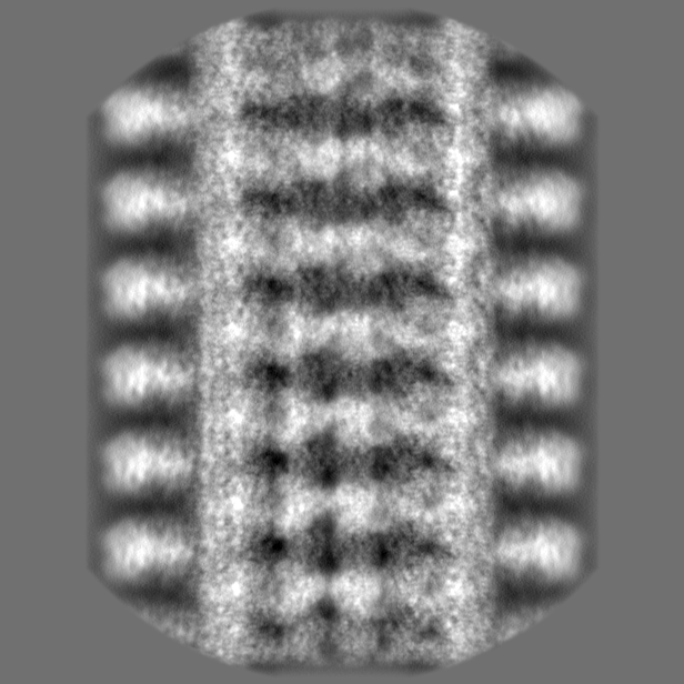
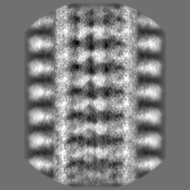
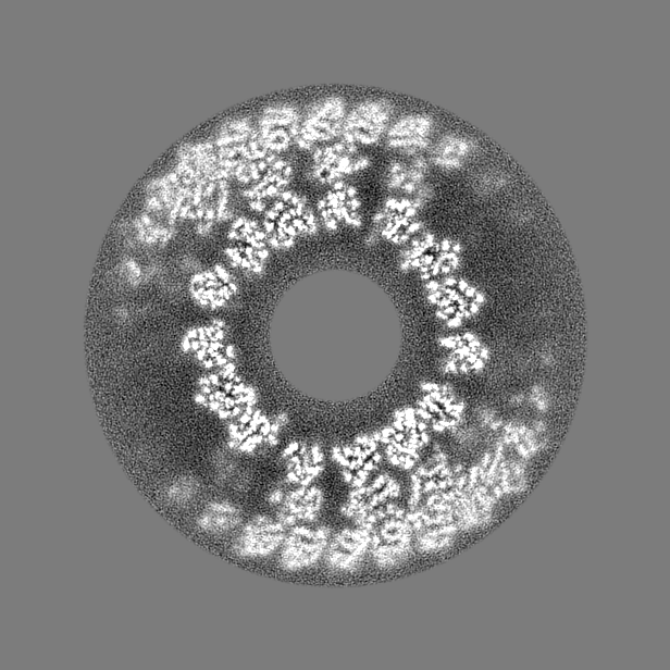
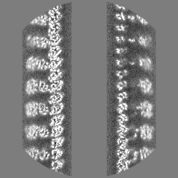
Surface view with section colored by density value
Name: microtubule / type: organelle_or_cellular_component / ID: 3 / Parent: 1 / Macromolecule list: #2, #1 Details: Chains A and B make up a curved microtubule protofilament.
Source (natural)
Organism: Sus scrofa (pig)
+
Macromolecule #1: Tubulin alpha-1B chain
Macromolecule
Name: Tubulin alpha-1B chain / type: protein_or_peptide / ID: 1 / Number of copies: 2 / Enantiomer: LEVO
In the structure databanks used in Yorodumi, some data are registered as the other names, "COVID-19 virus" and "2019-nCoV". Here are the details of the virus and the list of structure data.
Jan 31, 2019. EMDB accession codes are about to change! (news from PDBe EMDB page)
EMDB accession codes are about to change! (news from PDBe EMDB page)
The allocation of 4 digits for EMDB accession codes will soon come to an end. Whilst these codes will remain in use, new EMDB accession codes will include an additional digit and will expand incrementally as the available range of codes is exhausted. The current 4-digit format prefixed with “EMD-” (i.e. EMD-XXXX) will advance to a 5-digit format (i.e. EMD-XXXXX), and so on. It is currently estimated that the 4-digit codes will be depleted around Spring 2019, at which point the 5-digit format will come into force.
The EM Navigator/Yorodumi systems omit the EMD- prefix.
Related info.:Q: What is EMD? / ID/Accession-code notation in Yorodumi/EM Navigator
Yorodumi is a browser for structure data from EMDB, PDB, SASBDB, etc.
This page is also the successor to EM Navigator detail page, and also detail information page/front-end page for Omokage search.
The word "yorodu" (or yorozu) is an old Japanese word meaning "ten thousand". "mi" (miru) is to see.
Related info.:EMDB / PDB / SASBDB / Comparison of 3 databanks / Yorodumi Search / Aug 31, 2016. New EM Navigator & Yorodumi / Yorodumi Papers / Jmol/JSmol / Function and homology information / Changes in new EM Navigator and Yorodumi
 Movie
Movie Controller


 Open data
 Basic information
 Map data
 Sample
 Keywords
 Function and homology information

 Authors
 United States, 1 items
  Citation
 Structure visualization
 Downloads & links
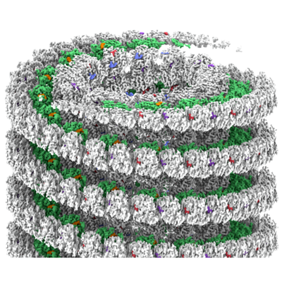 emd_7026.png
 http://ftp.pdbj.org/pub/emdb/structures/EMD-7026


 Z (Sec.)
 Y (Row.)
 X (Col.)


 Sample components


 Processing
 Electron microscopy
 FIELD EMISSION GUN