 Movie
Movie Controller


+ Open data

- Basic information
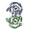
| Entry | Database: PDB / ID: 2btq | ||||||
|---|---|---|---|---|---|---|---|
| Title | Structure of BtubAB heterodimer from Prosthecobacter dejongeii | ||||||
 Components Components |
| ||||||
 Keywords Keywords | STRUCTURAL PROTEIN / CYTOSKELETAL PROTEIN-COMPLEX / BACTERIAL TUBULIN / CYTOSKELETON / POLYMERIZATION / VERRUCOMICROBIA / PROTEIN COMPLEX / CYTOSKELETAL PROTEIN | ||||||
| Function / homology |  Function and homology information Function and homology informationmicrotubule-based process / structural constituent of cytoskeleton / microtubule / hydrolase activity / GTPase activity / GTP binding Similarity search - Function | ||||||
| Biological species |  PROSTHECOBACTER DEJONGEII (bacteria) PROSTHECOBACTER DEJONGEII (bacteria) | ||||||
| Method |  X-RAY DIFFRACTION / SYNCHROTRON / MOLECULAR REPLACEMENT / Resolution: 3.2 Å X-RAY DIFFRACTION / SYNCHROTRON / MOLECULAR REPLACEMENT / Resolution: 3.2 Å | ||||||
 Authors Authors | Schlieper, D. / Lowe, J. | ||||||
 Citation Citation | Journal: Proc.Natl.Acad.Sci.USA / Year: 2005 Title: Structure of Bacterial Tubulin Btuba/B: Evidence for Horizontal Gene Transfer. Authors: Schlieper, D. / Oliva, M.A. / Andreu, J.M. / Lowe, J. | ||||||
| History |
|
- Structure visualization
| Structure viewer | Molecule: MolmilJmol/JSmol |
|---|
- Downloads & links
-Download
| PDBx/mmCIF format | 2btq.cif.gz | 172.5 KB | Display | PDBx/mmCIF format |
|---|---|---|---|---|
| PDB format | pdb2btq.ent.gz | 135.1 KB | Display | PDB format |
| PDBx/mmJSON format | 2btq.json.gz | Tree view | PDBx/mmJSON format | |
| Others |  Other downloads Other downloads |
-Validation report
| Arichive directory | https://data.pdbj.org/pub/pdb/validation_reports/bt/2btqftp://data.pdbj.org/pub/pdb/validation_reports/bt/2btq | HTTPS FTP |
|---|
-Related structure data
| Related structure data |  2btoSC S: Starting model for refinement C: citing same article ( |
|---|---|
| Similar structure data |


-Links
 PDBj
PDBj


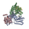
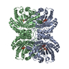
- Assembly
| Deposited unit | 
| ||||||||
|---|---|---|---|---|---|---|---|---|---|
| 1 | 
| ||||||||
| Unit cell |
|
-Components
| #1: Protein | Mass: 51299.371 Da / Num. of mol.: 1 Source method: isolated from a genetically manipulated source Source: (gene. exp.) PROSTHECOBACTER DEJONGEII (bacteria)Description: GERMAN COLLECTION OF MICROORGANISMS (DSM 12251) Plasmid: PHIS17 / Production host: | ||
|---|---|---|---|
| #2: Protein | Mass: 46465.508 Da / Num. of mol.: 1 Source method: isolated from a genetically manipulated source Source: (gene. exp.) PROSTHECOBACTER DEJONGEII (bacteria)Description: GERMAN COLLECTION OF MICROORGANISMS (DSM 12251) Plasmid: PHIS17 / Production host: | ||
| #3: Chemical | ChemComp-GDP /   Type: RNA linking / Mass: 443.201 Da / Num. of mol.: 1 / Source method: obtained synthetically / Formula: C10H15N5O11P2 / Comment: GDP, energy-carrying molecule*YM Type: RNA linking / Mass: 443.201 Da / Num. of mol.: 1 / Source method: obtained synthetically / Formula: C10H15N5O11P2 / Comment: GDP, energy-carrying molecule*YM | ||
| #4: Chemical | ChemComp-SO4 /   Mass: 96.063 Da / Num. of mol.: 4 / Source method: obtained synthetically / Formula: SO4 Mass: 96.063 Da / Num. of mol.: 4 / Source method: obtained synthetically / Formula: SO4Sequence details | THE SEQADV RECORDS BELOW FOR THE BTUBA SEQUENCE APPEARS TO BE FROM A NATURAL VARIATION IN THE GENE. | |
-Experimental details
-Experiment
| Experiment | Method: X-RAY DIFFRACTION / Number of used crystals: 1 |
|---|
- Sample preparation
| Crystal | Density Matthews: 4.51 Å3/Da / Density % sol: 72.73 % |
|---|---|
| Crystal grow | pH: 7.5 / Details: 1.5M LI2SO4, 0.4M TRIS/HCL, PH 7.5 |
-Data collection
| Diffraction | Mean temperature: 100 K |
|---|---|
| Diffraction source | Source: SYNCHROTRON / Site: ESRF  / Beamline: ID14-1 / Wavelength: 0.934 / Beamline: ID14-1 / Wavelength: 0.934 |
| Detector | Type: ADSC CCD / Detector: CCD / Date: Dec 8, 2004 |
| Radiation | Protocol: SINGLE WAVELENGTH / Monochromatic (M) / Laue (L): M / Scattering type: x-ray |
| Radiation wavelength | Wavelength: 0.934 Å / Relative weight: 1 |
| Reflection | Resolution: 3.2→53.52 Å / Num. obs: 29998 / % possible obs: 99 % / Observed criterion σ(I): 2.6 / Redundancy: 3.6 % / Rmerge(I) obs: 0.09 / Net I/σ(I): 12.8 |
| Reflection shell | Resolution: 3.2→3.37 Å / Redundancy: 3.6 % / Rmerge(I) obs: 0.38 / Mean I/σ(I) obs: 2.6 / % possible all: 99.5 |
- Processing
| Software |
| ||||||||||||||||||||||||||||||||||||||||||||||||||||||||||||
|---|---|---|---|---|---|---|---|---|---|---|---|---|---|---|---|---|---|---|---|---|---|---|---|---|---|---|---|---|---|---|---|---|---|---|---|---|---|---|---|---|---|---|---|---|---|---|---|---|---|---|---|---|---|---|---|---|---|---|---|---|---|
| Refinement | Method to determine structure: MOLECULAR REPLACEMENT Starting model: PDB ENTRY 2BTO Resolution: 3.2→53.52 Å / Data cutoff high absF: 10000 / Cross valid method: THROUGHOUT / σ(F): 0 Stereochemistry target values: MAXIMUM LIKELIHOOD TARGET USING AMPLITUDES
| ||||||||||||||||||||||||||||||||||||||||||||||||||||||||||||
| Solvent computation | Bsol: 90.9632 Å2 / ksol: 0.339518 e/Å3 | ||||||||||||||||||||||||||||||||||||||||||||||||||||||||||||
| Displacement parameters |
| ||||||||||||||||||||||||||||||||||||||||||||||||||||||||||||
| Refine analyze |
| ||||||||||||||||||||||||||||||||||||||||||||||||||||||||||||
| Refinement step | Cycle: LAST / Resolution: 3.2→53.52 Å
| ||||||||||||||||||||||||||||||||||||||||||||||||||||||||||||
| Refine LS restraints |
| ||||||||||||||||||||||||||||||||||||||||||||||||||||||||||||
| LS refinement shell | Resolution: 3.2→3.24 Å / Total num. of bins used: 30 /
| ||||||||||||||||||||||||||||||||||||||||||||||||||||||||||||
| Xplor file |
|
