 Movie
Movie Controller

[English] 日本語
 Yorodumi
- PDB-4df9: Crystal structure of a putative peptidase (BF3526) from Bacteroid... -
+ Open data

- Basic information
| Entry | Database: PDB / ID: 4df9 | ||||||
|---|---|---|---|---|---|---|---|
| Title | Crystal structure of a putative peptidase (BF3526) from Bacteroides fragilis NCTC 9343 at 2.17 A resolution | ||||||
 Components Components | putative peptidase | ||||||
 Keywords Keywords | HYDROLASE / IgA Peptidase M64 / Structural Genomics / Joint Center for Structural Genomics / JCSG / Protein Structure Initiative / PSI-BIOLOGY | ||||||
| Function / homology |  Function and homology information Function and homology information | ||||||
| Biological species |  Bacteroides fragilis NCTC 9343 (bacteria) Bacteroides fragilis NCTC 9343 (bacteria) | ||||||
| Method |  X-RAY DIFFRACTION / SYNCHROTRON / MAD / Resolution: 2.17 Å X-RAY DIFFRACTION / SYNCHROTRON / MAD / Resolution: 2.17 Å | ||||||
 Authors Authors | Joint Center for Structural Genomics (JCSG) | ||||||
 Citation Citation | Journal: To be published Title: Crystal structure of a putative peptidase (BF3526) from Bacteroides fragilis NCTC 9343 at 2.17 A resolution Authors: Joint Center for Structural Genomics (JCSG) | ||||||
| History |
|
- Structure visualization
| Structure viewer | Molecule: MolmilJmol/JSmol |
|---|
- Downloads & links
-Download
| PDBx/mmCIF format | 4df9.cif.gz | 1013.3 KB | Display | PDBx/mmCIF format |
|---|---|---|---|---|
| PDB format | pdb4df9.ent.gz | 840.5 KB | Display | PDB format |
| PDBx/mmJSON format | 4df9.json.gz | Tree view | PDBx/mmJSON format | |
| Others |  Other downloads Other downloads |
-Validation report
| Arichive directory | https://data.pdbj.org/pub/pdb/validation_reports/df/4df9ftp://data.pdbj.org/pub/pdb/validation_reports/df/4df9 | HTTPS FTP |
|---|
-Related structure data
| Similar structure data | |
|---|---|
| Other databases |


-Links
 PDBj
PDBj


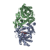
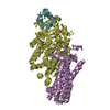
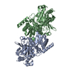
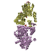
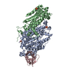
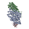
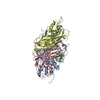
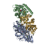
- Assembly
| Deposited unit | 
| ||||||||
|---|---|---|---|---|---|---|---|---|---|
| 1 | 
| ||||||||
| 2 | 
| ||||||||
| 3 | 
| ||||||||
| Unit cell |
| ||||||||
| Details | ANALYTICAL SIZE EXCLUSION CHROMATOGRAPHY PROVIDES SUPPORTING EVIDENCE THAT A DIMER IS A SIGNIFICANT OLIGOMERIC STATE IN SOLUTION. |
-Components
-Protein , 1 types, 6 molecules ABCDEF
| #1: Protein | Mass: 46430.191 Da / Num. of mol.: 6 Source method: isolated from a genetically manipulated source Source: (gene. exp.) Bacteroides fragilis NCTC 9343 (bacteria)Strain: NCTC 9343 / Gene: BF3526, BF9343_3433 / Plasmid: SpeedET / Production host: |
|---|
-Non-polymers , 7 types, 2022 molecules 


| #2: Chemical | ChemComp-NA /  Mass: 22.990 Da / Num. of mol.: 6 / Source method: obtained synthetically / Formula: Na Mass: 22.990 Da / Num. of mol.: 6 / Source method: obtained synthetically / Formula: Na#3: Chemical | ChemComp-ZN /  Mass: 65.409 Da / Num. of mol.: 12 / Source method: obtained synthetically / Formula: Zn Mass: 65.409 Da / Num. of mol.: 12 / Source method: obtained synthetically / Formula: Zn#4: Chemical | ChemComp-2PE /  Mass: 414.488 Da / Num. of mol.: 6 / Source method: obtained synthetically / Formula: C18H38O10 / Comment: precipitant*YM Mass: 414.488 Da / Num. of mol.: 6 / Source method: obtained synthetically / Formula: C18H38O10 / Comment: precipitant*YM#5: Chemical | ChemComp-PO4 /  Mass: 94.971 Da / Num. of mol.: 5 / Source method: obtained synthetically / Formula: PO4 Mass: 94.971 Da / Num. of mol.: 5 / Source method: obtained synthetically / Formula: PO4#6: Chemical | ChemComp-GOL /  Mass: 92.094 Da / Num. of mol.: 31 / Source method: obtained synthetically / Formula: C3H8O3 Mass: 92.094 Da / Num. of mol.: 31 / Source method: obtained synthetically / Formula: C3H8O3#7: Chemical | ChemComp-CL /  Mass: 35.453 Da / Num. of mol.: 15 / Source method: obtained synthetically / Formula: Cl Mass: 35.453 Da / Num. of mol.: 15 / Source method: obtained synthetically / Formula: Cl#8: Water | ChemComp-HOH / | Mass: 18.015 Da / Num. of mol.: 1947 / Source method: isolated from a natural source / Formula: H2O |
|---|
-Details
| Has protein modification | Y |
|---|---|
| Sequence details | THE CONSTRUCT (RESIDUES 19-426) WAS EXPRESSED WITH A PURIFICATION TAG MGSDKIHHHHHHENLYFQG. THE TAG ...THE CONSTRUCT (RESIDUES 19-426) WAS EXPRESSED WITH A PURIFICATI |
-Experimental details
-Experiment
| Experiment | Method: X-RAY DIFFRACTION / Number of used crystals: 1 |
|---|
- Sample preparation
| Crystal | Density Matthews: 2.82 Å3/Da / Density % sol: 56.44 % Description: DATA WERE SCALED USING XSCALE WITH FRIEDEL PAIRS KEPT AS SEPARATE WHEN COMPUTING R-SYM, COMPLETENESS AND Crystal grow | Temperature: 293 K / Method: vapor diffusion, sitting drop | Details: 20.00% Glycerol 20.00% polyethylene glycol 4000, NANODROP, VAPOR DIFFUSION, SITTING DROP, temperature 293K |
|---|
-Data collection
| Diffraction | Mean temperature: 100 K | |||||||||||||||||||||||||||||||||||||||||||||||||||||||||||||||||||||||||||||
|---|---|---|---|---|---|---|---|---|---|---|---|---|---|---|---|---|---|---|---|---|---|---|---|---|---|---|---|---|---|---|---|---|---|---|---|---|---|---|---|---|---|---|---|---|---|---|---|---|---|---|---|---|---|---|---|---|---|---|---|---|---|---|---|---|---|---|---|---|---|---|---|---|---|---|---|---|---|---|
| Diffraction source | Source: SYNCHROTRON / Site: SSRL  / Beamline: BL11-1 / Wavelength: 0.91837,0.97941,0.97899 / Beamline: BL11-1 / Wavelength: 0.91837,0.97941,0.97899 | |||||||||||||||||||||||||||||||||||||||||||||||||||||||||||||||||||||||||||||
| Detector | Type: MARMOSAIC 325 mm CCD / Detector: CCD / Date: Jul 22, 2011 Details: Flat mirror (vertical focusing); single crystal Si(111) bent monochromator (ho rizontal focusing) | |||||||||||||||||||||||||||||||||||||||||||||||||||||||||||||||||||||||||||||
| Radiation | Monochromator: single crystal Si(111) bent / Protocol: MAD / Monochromatic (M) / Laue (L): M / Scattering type: x-ray | |||||||||||||||||||||||||||||||||||||||||||||||||||||||||||||||||||||||||||||
| Radiation wavelength |
| |||||||||||||||||||||||||||||||||||||||||||||||||||||||||||||||||||||||||||||
| Reflection | Resolution: 2.17→48.732 Å / Num. obs: 162812 / % possible obs: 99 % / Observed criterion σ(I): -3 / Biso Wilson estimate: 32.817 Å2 / Rmerge(I) obs: 0.11 / Net I/σ(I): 10.11 | |||||||||||||||||||||||||||||||||||||||||||||||||||||||||||||||||||||||||||||
| Reflection shell | Diffraction-ID: 1
|
-Phasing
| Phasing | Method: MAD |
|---|
- Processing
| Software |
| |||||||||||||||||||||||||||||||||||||||||||||||||||||||||||||||||||||||||||||||||||||||||||||||||||||||||||||||||||||||||||||||||||||||||||||||||||||||||||||||||||||||||||||||
|---|---|---|---|---|---|---|---|---|---|---|---|---|---|---|---|---|---|---|---|---|---|---|---|---|---|---|---|---|---|---|---|---|---|---|---|---|---|---|---|---|---|---|---|---|---|---|---|---|---|---|---|---|---|---|---|---|---|---|---|---|---|---|---|---|---|---|---|---|---|---|---|---|---|---|---|---|---|---|---|---|---|---|---|---|---|---|---|---|---|---|---|---|---|---|---|---|---|---|---|---|---|---|---|---|---|---|---|---|---|---|---|---|---|---|---|---|---|---|---|---|---|---|---|---|---|---|---|---|---|---|---|---|---|---|---|---|---|---|---|---|---|---|---|---|---|---|---|---|---|---|---|---|---|---|---|---|---|---|---|---|---|---|---|---|---|---|---|---|---|---|---|---|---|---|---|---|
| Refinement | Method to determine structure: MAD / Resolution: 2.17→48.732 Å / Cor.coef. Fo:Fc: 0.958 / Cor.coef. Fo:Fc free: 0.9448 / Occupancy max: 1 / Occupancy min: 0.23 / Cross valid method: THROUGHOUT / σ(F): 0 Details: 1. A MET-INHIBITION PROTOCOL WAS USED FOR SELENOMETHIONINE INCORPORATION DURING PROTEIN EXPRESSION. THE OCCUPANCY OF THE SE ATOMS IN THE MSE RESIDUES WAS REDUCED TO 0.75 FOR THE REDUCED ...Details: 1. A MET-INHIBITION PROTOCOL WAS USED FOR SELENOMETHIONINE INCORPORATION DURING PROTEIN EXPRESSION. THE OCCUPANCY OF THE SE ATOMS IN THE MSE RESIDUES WAS REDUCED TO 0.75 FOR THE REDUCED SCATTERING POWER DUE TO PARTIAL S-MET INCORPORATION. 2. PHOSPHATE, POLYETHENE GLYCOL, GLYCEROL, SODIUM AND CHLORIDE MODELED ARE PRESENT IN THE CRYSTALLIZATION/CRYO/PROTEIN BUFFER CONDITIONS. 3. THE MODELING OF THE ZINC (ZN) IONS IS SUPPORTED BY PEAKS IN THE ANOMALOUS DIFFERENCE FOURIER MAPS AND ZINC K EMISSION LINES IN X-RAY FLUORESCENCE EXCITATION SPECTRA. 4. ATOM RECORD CONTAINS SUM OF TLS AND RESIDUAL B FACTORS. ANISOU RECORD CONTAINS SUM OF TLS AND RESIDUAL U FACTORS. 5. THE MAD PHASES WERE USED AS RESTRAINTS DURING REFINEMENT. 6. NCS RESTRAINTS WERE APPLIED USING BUSTER'S LSSR RESTRAINT REPRESENTATION (-AUTONCS)
| |||||||||||||||||||||||||||||||||||||||||||||||||||||||||||||||||||||||||||||||||||||||||||||||||||||||||||||||||||||||||||||||||||||||||||||||||||||||||||||||||||||||||||||||
| Displacement parameters | Biso max: 140.75 Å2 / Biso mean: 37.341 Å2 / Biso min: 4.16 Å2
| |||||||||||||||||||||||||||||||||||||||||||||||||||||||||||||||||||||||||||||||||||||||||||||||||||||||||||||||||||||||||||||||||||||||||||||||||||||||||||||||||||||||||||||||
| Refine analyze | Luzzati coordinate error obs: 0.226 Å | |||||||||||||||||||||||||||||||||||||||||||||||||||||||||||||||||||||||||||||||||||||||||||||||||||||||||||||||||||||||||||||||||||||||||||||||||||||||||||||||||||||||||||||||
| Refinement step | Cycle: LAST / Resolution: 2.17→48.732 Å
| |||||||||||||||||||||||||||||||||||||||||||||||||||||||||||||||||||||||||||||||||||||||||||||||||||||||||||||||||||||||||||||||||||||||||||||||||||||||||||||||||||||||||||||||
| Refine LS restraints |
| |||||||||||||||||||||||||||||||||||||||||||||||||||||||||||||||||||||||||||||||||||||||||||||||||||||||||||||||||||||||||||||||||||||||||||||||||||||||||||||||||||||||||||||||
| LS refinement shell | Resolution: 2.17→2.23 Å / Total num. of bins used: 20
| |||||||||||||||||||||||||||||||||||||||||||||||||||||||||||||||||||||||||||||||||||||||||||||||||||||||||||||||||||||||||||||||||||||||||||||||||||||||||||||||||||||||||||||||
| Refinement TLS params. | Method: refined / Refine-ID: X-RAY DIFFRACTION
| |||||||||||||||||||||||||||||||||||||||||||||||||||||||||||||||||||||||||||||||||||||||||||||||||||||||||||||||||||||||||||||||||||||||||||||||||||||||||||||||||||||||||||||||
| Refinement TLS group |
|
