 Movie
Movie Controller

[English] 日本語
 Yorodumi
- PDB-1x9j: Structure of butyrate kinase 2 reveals both open- and citrate-ind... -
+ Open data

- Basic information
| Entry | Database: PDB / ID: 1x9j | ||||||
|---|---|---|---|---|---|---|---|
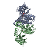
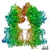
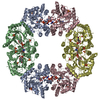
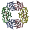
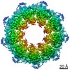
| Title | Structure of butyrate kinase 2 reveals both open- and citrate-induced closed conformations: implications for substrate-induced fit conformational changes | ||||||
 Components Components | Probable butyrate kinase 2 | ||||||
 Keywords Keywords | TRANSFERASE / ASKHA (ACETATE AND SUGAR KINASES / HSC70 / ACTIN) SUPERFAMILY / BUTYRATE KINASE / ACETATE KINASE / ISOBUTYRATE KINASE / TWO SIMILAR DOMAINS / BUTYRATE / ISOBUTYRATE / ENZYME MECHANISM | ||||||
| Function / homology |  Function and homology information Function and homology informationbutyrate kinase / butyrate kinase activity / acetate kinase activity / acetate metabolic process / ATP binding / cytoplasm Similarity search - Function | ||||||
| Biological species |   Thermotoga maritima (bacteria) Thermotoga maritima (bacteria) | ||||||
| Method |  X-RAY DIFFRACTION / SYNCHROTRON / MOLECULAR REPLACEMENT / Resolution: 3 Å X-RAY DIFFRACTION / SYNCHROTRON / MOLECULAR REPLACEMENT / Resolution: 3 Å | ||||||
 Authors Authors | Diao, J.S. / Sanders, D.A. / Hasson, M.S. | ||||||
 Citation Citation | Journal: To be Published Title: Structure of butyrate kinase 2 reveals both open and closed conformations of the two domains: implications for substrate-induced changes Authors: Diao, J.S. / Bhattacharyya, S. / Ma, Y.L.D. / Sanders, D.A. / Hasson, M.S. #1: Journal: Acta Crystallogr.,Sect.D / Year: 2003Title: Crystallization of Butyrate Kinase 2 from Thermotoga Maritima Mediated by Vapour Diffusion of Acetic Acid Authors: Diao, J.S. / Cooper, D.R. / Sanders, D.A. / Hasson, M.S. #2: Journal: To be PublishedTitle: Membership in the Askha Superfamily: Enzymological Properties and Crystal Structure of Butyrate Kinase 2 from Thermotoga Maritima Authors: Diao, J.S. / Cooper, D.R. / Bhattacharyya, S. / Sanders, D.A. / Hasson, M.S. | ||||||
| History |
|
- Structure visualization
| Structure viewer | Molecule: MolmilJmol/JSmol |
|---|
- Downloads & links
-Download
| PDBx/mmCIF format | 1x9j.cif.gz | 570.8 KB | Display | PDBx/mmCIF format |
|---|---|---|---|---|
| PDB format | pdb1x9j.ent.gz | 475.7 KB | Display | PDB format |
| PDBx/mmJSON format | 1x9j.json.gz | Tree view | PDBx/mmJSON format | |
| Others |  Other downloads Other downloads |
-Validation report
| Arichive directory | https://data.pdbj.org/pub/pdb/validation_reports/x9/1x9jftp://data.pdbj.org/pub/pdb/validation_reports/x9/1x9j | HTTPS FTP |
|---|
-Related structure data
| Related structure data |  1sazS S: Starting model for refinement |
|---|---|
| Similar structure data |
-Links
 PDBj
PDBj- Assembly
| Deposited unit | 
| ||||||||
|---|---|---|---|---|---|---|---|---|---|
| 1 |
| ||||||||
| 2 | 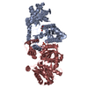
| ||||||||
| 3 | 
| ||||||||
| 4 | 
| ||||||||
| 5 | 
| ||||||||
| Unit cell |
| ||||||||
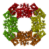
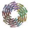
| Details | THE BIOLOGICAL UNIT IS IDENTICAL TO THE ASYMMETRIC UNIT, WHICH IS AN OCTAMER. |
-Components
-Protein , 1 types, 8 molecules ABCDEFGH
| #1: Protein | Mass: 42083.355 Da / Num. of mol.: 8 Source method: isolated from a genetically manipulated source Source: (gene. exp.) Thermotoga maritima (bacteria) / Gene: buk2 / Plasmid: PET30A(+) / Production host: |
|---|
-Non-polymers , 5 types, 62 molecules 


| #2: Chemical | ChemComp-GOL /  Mass: 92.094 Da / Num. of mol.: 5 / Source method: obtained synthetically / Formula: C3H8O3 Mass: 92.094 Da / Num. of mol.: 5 / Source method: obtained synthetically / Formula: C3H8O3#3: Chemical | ChemComp-PO4 /  Mass: 94.971 Da / Num. of mol.: 5 / Source method: obtained synthetically / Formula: PO4 Mass: 94.971 Da / Num. of mol.: 5 / Source method: obtained synthetically / Formula: PO4#4: Chemical | ChemComp-NA / |  Mass: 22.990 Da / Num. of mol.: 1 / Source method: obtained synthetically / Formula: Na Mass: 22.990 Da / Num. of mol.: 1 / Source method: obtained synthetically / Formula: Na#5: Chemical | ChemComp-CIT /  Mass: 192.124 Da / Num. of mol.: 4 / Source method: obtained synthetically / Formula: C6H8O7 Mass: 192.124 Da / Num. of mol.: 4 / Source method: obtained synthetically / Formula: C6H8O7#6: Water | ChemComp-HOH / | Mass: 18.015 Da / Num. of mol.: 47 / Source method: isolated from a natural source / Formula: H2O |
|---|
-Experimental details
-Experiment
| Experiment | Method: X-RAY DIFFRACTION / Number of used crystals: 1 |
|---|
- Sample preparation
| Crystal | Density Matthews: 3.6 Å3/Da / Density % sol: 65 % |
|---|---|
| Crystal grow | Temperature: 310 K / Method: vapor diffusion, sitting drop / pH: 4.2 Details: CRYSTALLIZATION CONDITIONS: THE PROTEIN SOLUTION CONTAINS 22.5-25 mg/ml BUK2, 25 mM TRIS-HCl pH 8.5, 400 mM NACl, 5 mM DTT, 10%(w/v) GLYCEROL. THE RESERVOIR SOLUTION IS 1ml of 77 mM ...Details: CRYSTALLIZATION CONDITIONS: THE PROTEIN SOLUTION CONTAINS 22.5-25 mg/ml BUK2, 25 mM TRIS-HCl pH 8.5, 400 mM NACl, 5 mM DTT, 10%(w/v) GLYCEROL. THE RESERVOIR SOLUTION IS 1ml of 77 mM PHOSPHATE-CITRATE AND 55% (w/v) PEG 200, pH 4.2, VAPOR DIFFUSION, SITTING DROP, temperature 310K |
-Data collection
| Diffraction | Mean temperature: 100 K |
|---|---|
| Diffraction source | Source: SYNCHROTRON / Site: APS  / Beamline: 14-ID-B / Wavelength: 0.9793 / Wavelength: 0.9793 Å / Beamline: 14-ID-B / Wavelength: 0.9793 / Wavelength: 0.9793 Å |
| Detector | Type: MAR scanner 345 mm plate / Detector: IMAGE PLATE / Date: Apr 27, 2002 / Details: MIRRORS |
| Radiation | Protocol: SINGLE WAVELENGTH / Monochromatic (M) / Laue (L): M / Scattering type: x-ray |
| Radiation wavelength | Wavelength: 0.9793 Å / Relative weight: 1 |
| Reflection | Resolution: 3→100 Å / Num. all: 88518 / Num. obs: 88350 / % possible obs: 97 % / Redundancy: 14.2 % / Biso Wilson estimate: 68.8 Å2 / Rsym value: 0.093 / Net I/σ(I): 11.9 |
| Reflection shell | Resolution: 3→3.11 Å / Redundancy: 5.6 % / Mean I/σ(I) obs: 4.2 / Num. unique all: 7233 / Rsym value: 0.359 / % possible all: 80.3 |
- Processing
| Software |
| ||||||||||||||||||||||||||||||||||||
|---|---|---|---|---|---|---|---|---|---|---|---|---|---|---|---|---|---|---|---|---|---|---|---|---|---|---|---|---|---|---|---|---|---|---|---|---|---|
| Refinement | Method to determine structure: MOLECULAR REPLACEMENT Starting model: PDB entry 1SAZ Resolution: 3→91.48 Å / Rfactor Rfree error: 0.003 / Data cutoff high absF: 3022342.25 / Data cutoff low absF: 0 / Isotropic thermal model: ISOROPIC / Cross valid method: THROUGHOUT / σ(F): 2 / Stereochemistry target values: Engh & Huber
| ||||||||||||||||||||||||||||||||||||
| Solvent computation | Solvent model: FLAT MODEL / Bsol: 37.0944 Å2 / ksol: 0.346204 e/Å3 | ||||||||||||||||||||||||||||||||||||
| Displacement parameters | Biso mean: 55.2 Å2 | ||||||||||||||||||||||||||||||||||||
| Refine analyze |
| ||||||||||||||||||||||||||||||||||||
| Refinement step | Cycle: LAST / Resolution: 3→91.48 Å
| ||||||||||||||||||||||||||||||||||||
| Refine LS restraints |
| ||||||||||||||||||||||||||||||||||||
| Refine LS restraints NCS | NCS model details: RESTRAINTS | ||||||||||||||||||||||||||||||||||||
| LS refinement shell | Resolution: 3→3.11 Å / Rfactor Rfree error: 0.015 / Total num. of bins used: 10
| ||||||||||||||||||||||||||||||||||||
| Xplor file |
|
