 Movie
Movie Controller

[English] 日本語
 Yorodumi
- PDB-1jbz: CRYSTAL STRUCTURE ANALYSIS OF A DUAL-WAVELENGTH EMISSION GREEN FL... -
+ Open data

- Basic information
| Entry | Database: PDB / ID: 1jbz | ||||||
|---|---|---|---|---|---|---|---|
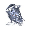
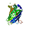
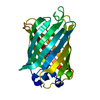
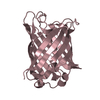
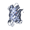
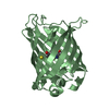
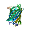
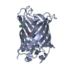
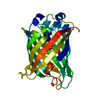
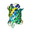
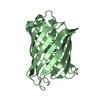
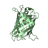
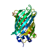
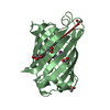
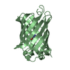
| Title | CRYSTAL STRUCTURE ANALYSIS OF A DUAL-WAVELENGTH EMISSION GREEN FLUORESCENT PROTEIN VARIANT AT HIGH PH | ||||||
 Components Components | GREEN FLUORESCENT PROTEIN | ||||||
 Keywords Keywords | LUMINESCENT PROTEIN / Beta Barrel / Chromophore | ||||||
| Function / homology |  Function and homology information Function and homology information | ||||||
| Biological species |   Aequorea victoria (jellyfish) Aequorea victoria (jellyfish) | ||||||
| Method |  X-RAY DIFFRACTION / SYNCHROTRON / MOLECULAR REPLACEMENT / Resolution: 1.5 Å X-RAY DIFFRACTION / SYNCHROTRON / MOLECULAR REPLACEMENT / Resolution: 1.5 Å | ||||||
 Authors Authors | Hanson, G.T. / McAnaney, T.B. / Park, E.S. / Rendell, M.E.P. / Yarbrough, D.K. / Chu, S. / Xi, L. / Boxer, S.G. / Montrose, M.H. / Remington, S.J. | ||||||
 Citation Citation | Journal: Biochemistry / Year: 2002 Title: Green Fluorescent Protein Variants as Ratiometric Dual Emission pH Sensors. 1. Structural Characterization and Preliminary Application. Authors: Hanson, G.T. / McAnaney, T.B. / Park, E.S. / Rendell, M.E.P. / Yarbrough, D.K. / Chu, S. / Xi, L. / Boxer, S.G. / Montrose, M.H. / Remington, S.J. | ||||||
| History |
|
- Structure visualization
| Structure viewer | Molecule: MolmilJmol/JSmol |
|---|
- Downloads & links
-Download
| PDBx/mmCIF format | 1jbz.cif.gz | 64.9 KB | Display | PDBx/mmCIF format |
|---|---|---|---|---|
| PDB format | pdb1jbz.ent.gz | 45.4 KB | Display | PDB format |
| PDBx/mmJSON format | 1jbz.json.gz | Tree view | PDBx/mmJSON format | |
| Others |  Other downloads Other downloads |
-Validation report
| Summary document | 1jbz_validation.pdf.gz | 432.6 KB | Display | wwPDB validaton report |
|---|---|---|---|---|
| Full document | 1jbz_full_validation.pdf.gz | 438.1 KB | Display | |
| Data in XML | 1jbz_validation.xml.gz | 13.5 KB | Display | |
| Data in CIF | 1jbz_validation.cif.gz | 19.4 KB | Display | |
| Arichive directory | https://data.pdbj.org/pub/pdb/validation_reports/jb/1jbzftp://data.pdbj.org/pub/pdb/validation_reports/jb/1jbz | HTTPS FTP |
-Related structure data
| Related structure data |  1jbyC  1emaS C: citing same article ( S: Starting model for refinement |
|---|---|
| Similar structure data |


-Links
 PDBj
PDBj

- Assembly
| Deposited unit | 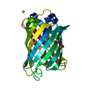
| ||||||||
|---|---|---|---|---|---|---|---|---|---|
| 1 |
| ||||||||
| Unit cell |
|
-Components
| #1: Protein | Mass: 26866.326 Da / Num. of mol.: 1 / Mutation: S65T,Q80R,H148G,T203C Source method: isolated from a genetically manipulated source Source: (gene. exp.) Aequorea victoria (jellyfish) / Plasmid: pRSETb / Production host:  | ||||||||
|---|---|---|---|---|---|---|---|---|---|
| #2: Chemical |   Mass: 24.305 Da / Num. of mol.: 2 / Source method: obtained synthetically / Formula: Mg Mass: 24.305 Da / Num. of mol.: 2 / Source method: obtained synthetically / Formula: Mg#3: Chemical |   Mass: 62.068 Da / Num. of mol.: 3 / Source method: obtained synthetically / Formula: C2H6O2 Mass: 62.068 Da / Num. of mol.: 3 / Source method: obtained synthetically / Formula: C2H6O2#4: Water | ChemComp-HOH / |  Mass: 18.015 Da / Num. of mol.: 202 / Source method: isolated from a natural source / Formula: H2O Mass: 18.015 Da / Num. of mol.: 202 / Source method: isolated from a natural source / Formula: H2OHas protein modification | Y | Sequence details | RESIDUES 65, 66, AND 67 ARE NOT PRESENT IN THE ENTRY AND ARE INSTEAD REPLACED WITH CRO 66 | |
-Experimental details
-Experiment
| Experiment | Method: X-RAY DIFFRACTION / Number of used crystals: 1 |
|---|
- Sample preparation
| Crystal | Density Matthews: 2.05 Å3/Da / Density % sol: 40 % | ||||||||||||||||||||||||||||||||||||||||||
|---|---|---|---|---|---|---|---|---|---|---|---|---|---|---|---|---|---|---|---|---|---|---|---|---|---|---|---|---|---|---|---|---|---|---|---|---|---|---|---|---|---|---|---|
| Crystal grow | Temperature: 295 K / Method: vapor diffusion, hanging drop / pH: 9 Details: PEG 4000, Tris, Magnesium chloride, pH 9.0, VAPOR DIFFUSION, HANGING DROP, temperature 295K | ||||||||||||||||||||||||||||||||||||||||||
| Crystal grow | *PLUS pH: 7.9 | ||||||||||||||||||||||||||||||||||||||||||
| Components of the solutions | *PLUS
|
-Data collection
| Diffraction | Mean temperature: 100 K |
|---|---|
| Diffraction source | Source: SYNCHROTRON / Site: SSRL  / Beamline: BL7-1 / Wavelength: 1.08 Å / Beamline: BL7-1 / Wavelength: 1.08 Å |
| Detector | Type: MAR scanner 345 mm plate / Detector: IMAGE PLATE / Date: Nov 27, 2000 Details: 58 cm long, Pt-coated fused silica, vertical focus mirror |
| Radiation | Monochromator: Cylindrically bent triangular Si(111) asymmetric cut horizontal focus Protocol: SINGLE WAVELENGTH / Monochromatic (M) / Laue (L): M / Scattering type: x-ray |
| Radiation wavelength | Wavelength: 1.08 Å / Relative weight: 1 |
| Reflection | Resolution: 1.5→19.9 Å / Num. all: 120478 / Num. obs: 34536 / % possible obs: 96.2 % / Redundancy: 3.5 % / Biso Wilson estimate: 16.6 Å2 / Rmerge(I) obs: 0.045 / Net I/σ(I): 9.3 |
| Reflection shell | Resolution: 1.5→1.57 Å / Redundancy: 3.3 % / Rmerge(I) obs: 0.319 / Mean I/σ(I) obs: 2.3 / Num. unique all: 4801 / % possible all: 93.6 |
| Reflection | *PLUS Num. measured all: 120478 |
| Reflection shell | *PLUS Lowest resolution: 1.58 Å / % possible obs: 93.6 % |
- Processing
| Software |
| ||||||||||||||||||
|---|---|---|---|---|---|---|---|---|---|---|---|---|---|---|---|---|---|---|---|
| Refinement | Method to determine structure: MOLECULAR REPLACEMENT Starting model: PDB ENTRY 1EMA Resolution: 1.5→19.9 Å / Stereochemistry target values: ENGH & HUBER /
| ||||||||||||||||||
| Refinement step | Cycle: LAST / Resolution: 1.5→19.9 Å
| ||||||||||||||||||
| Refine LS restraints |
| ||||||||||||||||||
| LS refinement shell | Resolution: 1.5→1.57 Å /
| ||||||||||||||||||
| Refinement | *PLUS Rfactor all: 0.177 | ||||||||||||||||||
| Solvent computation | *PLUS | ||||||||||||||||||
| Displacement parameters | *PLUS | ||||||||||||||||||
| Refine LS restraints | *PLUS
|
