 Movie
Movie Controller

[English] 日本語
 Yorodumi
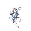
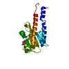
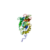
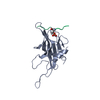
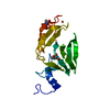
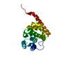
- PDB-1j4l: SOLUTION STRUCTURE OF THE FHA2 DOMAIN OF RAD53 COMPLEXED WITH A P... -
+ Open data

- Basic information
| Entry | Database: PDB / ID: 1j4l | ||||||
|---|---|---|---|---|---|---|---|
| Title | SOLUTION STRUCTURE OF THE FHA2 DOMAIN OF RAD53 COMPLEXED WITH A PHOSPHOTHREONYL PEPTIDE DERIVED FROM RAD9 | ||||||
 Components Components |
| ||||||
 Keywords Keywords | TRANSFERASE / FHA DOMAIN / RAD53 / RAD9 / PHOSPHOTHREONINE / PHOSPHOPROTEIN | ||||||
| Function / homology |  Function and homology information Function and homology informationG2/M DNA damage checkpoint / Chk1/Chk2(Cds1) mediated inactivation of Cyclin B:Cdk1 complex / deoxyribonucleoside triphosphate biosynthetic process / negative regulation of DNA strand resection involved in replication fork processing / mitotic DNA damage checkpoint signaling / negative regulation of phosphorylation / SUMOylation of transcription factors / dual-specificity kinase / Recruitment and ATM-mediated phosphorylation of repair and signaling proteins at DNA double strand breaks / mitotic intra-S DNA damage checkpoint signaling ...G2/M DNA damage checkpoint / Chk1/Chk2(Cds1) mediated inactivation of Cyclin B:Cdk1 complex / deoxyribonucleoside triphosphate biosynthetic process / negative regulation of DNA strand resection involved in replication fork processing / mitotic DNA damage checkpoint signaling / negative regulation of phosphorylation / SUMOylation of transcription factors / dual-specificity kinase / Recruitment and ATM-mediated phosphorylation of repair and signaling proteins at DNA double strand breaks / mitotic intra-S DNA damage checkpoint signaling / Ubiquitin-Mediated Degradation of Phosphorylated Cdc25A / telomere maintenance in response to DNA damage / negative regulation of DNA damage checkpoint / DNA replication origin binding / DNA replication initiation / regulation of DNA repair / site of DNA damage / mitotic G1 DNA damage checkpoint signaling / protein serine/threonine/tyrosine kinase activity / DNA damage checkpoint signaling / nucleotide-excision repair / enzyme activator activity / intracellular protein localization / double-strand break repair / protein tyrosine kinase activity / double-stranded DNA binding / histone binding / protein kinase activity / regulation of cell cycle / protein serine kinase activity / DNA repair / protein serine/threonine kinase activity / chromatin / positive regulation of transcription by RNA polymerase II / ATP binding / nucleus / cytosol / cytoplasm Similarity search - Function | ||||||
| Biological species |  | ||||||
| Method | SOLUTION NMR / THE COMPLEX STRUCTURES ARE GENERATED USING A TOTAL OF 3369 RESTRAINTS, 3181 DISTANCE RESTRAINTS, AND 188 TALOS-DERIVED DIHEDRAL ANGLE RESTRAINTS. | ||||||
 Authors Authors | Byeon, I.-J.L. / Yongkiettrakul, S. / Tsai, M.-D. | ||||||
 Citation Citation | Journal: J.Mol.Biol. / Year: 2001 Title: Solution structure of the yeast Rad53 FHA2 complexed with a phosphothreonine peptide pTXXL: comparison with the structures of FHA2-pYXL and FHA1-pTXXD complexes. Authors: Byeon, I.J. / Yongkiettrakul, S. / Tsai, M.D. #1: Journal: J.Mol.Biol. / Year: 2000Title: II. Structure and Specificity of the Interaction between the Fha2 Domain of Rad53 and Phosphotyrosyl Peptides. Authors: Wang, P. / Byeon, I.J. / Liao, H. / Beebe, K.D. / Yongkiettrakul, S. / Pei, D. / Tsai, M.D. #2: Journal: J.Mol.Biol. / Year: 1999Title: Structure and Function of a New Phosphopeptide-Binding Domain Containing the Fha2 of Rad53. Authors: Liao, H. / Byeon, I.J. / Tsai, M.D. | ||||||
| History |
|
- Structure visualization
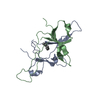
| Structure viewer | Molecule: MolmilJmol/JSmol |
|---|
- Downloads & links
-Download
| PDBx/mmCIF format | 1j4l.cif.gz | 72.1 KB | Display | PDBx/mmCIF format |
|---|---|---|---|---|
| PDB format | pdb1j4l.ent.gz | 53.7 KB | Display | PDB format |
| PDBx/mmJSON format | 1j4l.json.gz | Tree view | PDBx/mmJSON format | |
| Others |  Other downloads Other downloads |
-Validation report
| Arichive directory | https://data.pdbj.org/pub/pdb/validation_reports/j4/1j4lftp://data.pdbj.org/pub/pdb/validation_reports/j4/1j4l | HTTPS FTP |
|---|
-Related structure data


-Links
 PDBj
PDBj


- Assembly
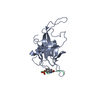
| Deposited unit | 
| |||||||||
|---|---|---|---|---|---|---|---|---|---|---|
| 1 |
| |||||||||
| NMR ensembles |
|
-Components
| #1: Protein | Mass: 18148.758 Da / Num. of mol.: 1 / Fragment: C-TERMINAL FHA DOMAIN (FHA2) Source method: isolated from a genetically manipulated source Source: (gene. exp.) Gene: SPK1 OR RAD53 / Plasmid: PGEX-4T / Species (production host): Escherichia coli / Production host:  References: UniProt: P22216, Transferases; Transferring phosphorus-containing groups; Phosphotransferases with an alcohol group as acceptor |
|---|---|
| #2: Protein/peptide | Mass: 1137.130 Da / Num. of mol.: 1 / Fragment: RESIDUES 599-607 / Source method: obtained synthetically Details: THIS PHOSPHOTHREONYL PEPTIDE WAS CHEMICALLY SYNTHESIZED. References: UniProt: P14737 |
| Has protein modification | Y |
-Experimental details
-Experiment
| Experiment | Method: SOLUTION NMR | ||||||||||||||||||||
|---|---|---|---|---|---|---|---|---|---|---|---|---|---|---|---|---|---|---|---|---|---|
| NMR experiment |
| ||||||||||||||||||||
| NMR details | Text: THE STRUCTURE WAS DETERMINED USING TRIPLE- RESONANCE NMR SPECTROSCOPY. |
- Sample preparation
| Details | Contents: 0.5 MM FHA2 U-15N,13C 1.5 MM PHOSPHOTHREONYL PEPTIDE OF RAD9; 10 MM SODIUM PHOSPHATE(PH 6.5), 1 MM DTT, AND 1 MM EDTA |
|---|---|
| Sample conditions | Ionic strength: 10 mM SODIUM PHOSPHATE, 1 mM DTT, AND 1 mM EDTA pH: 6.5 / Pressure: AMBIENT / Temperature: 293.00 K |
| Crystal grow | *PLUS Method: other / Details: NMR |
-NMR measurement
| NMR spectrometer | Type: Bruker DRX / Manufacturer: Bruker / Model: DRX / Field strength: 800 MHz |
|---|
- Processing
| NMR software |
| ||||||||||||
|---|---|---|---|---|---|---|---|---|---|---|---|---|---|
| Refinement | Method: THE COMPLEX STRUCTURES ARE GENERATED USING A TOTAL OF 3369 RESTRAINTS, 3181 DISTANCE RESTRAINTS, AND 188 TALOS-DERIVED DIHEDRAL ANGLE RESTRAINTS. Software ordinal: 1 | ||||||||||||
| NMR ensemble | Conformers submitted total number: 1 |
 X-PLOR