 Movie
Movie Controller

[English] 日本語
 Yorodumi
- PDB-1k2m: Solution Structure of the FHA2 Domain of Rad53 Complexed with a P... -
+ Open data

- Basic information
| Entry | Database: PDB / ID: 1k2m | |||||||||
|---|---|---|---|---|---|---|---|---|---|---|
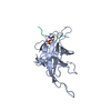
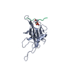
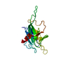
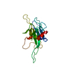
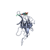
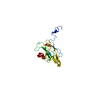
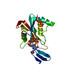
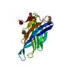
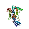
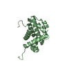
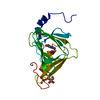
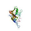
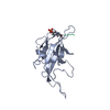
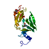
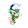
| Title | Solution Structure of the FHA2 Domain of Rad53 Complexed with a Phosphotyrosyl Peptide Derived from Rad9 | |||||||||
 Components Components |
| |||||||||
 Keywords Keywords | TRANSFERASE / FHA domain / Rad53 / Rad9 / Phosphotyrosine / phosphoprotein | |||||||||
| Function / homology |  Function and homology information Function and homology informationdeoxyribonucleoside triphosphate biosynthetic process / negative regulation of DNA strand resection involved in replication fork processing / meiotic recombination checkpoint signaling / SUMOylation of transcription factors / dual-specificity kinase / Recruitment and ATM-mediated phosphorylation of repair and signaling proteins at DNA double strand breaks / mitotic intra-S DNA damage checkpoint signaling / telomere maintenance in response to DNA damage / negative regulation of DNA damage checkpoint / DNA replication origin binding ...deoxyribonucleoside triphosphate biosynthetic process / negative regulation of DNA strand resection involved in replication fork processing / meiotic recombination checkpoint signaling / SUMOylation of transcription factors / dual-specificity kinase / Recruitment and ATM-mediated phosphorylation of repair and signaling proteins at DNA double strand breaks / mitotic intra-S DNA damage checkpoint signaling / telomere maintenance in response to DNA damage / negative regulation of DNA damage checkpoint / DNA replication origin binding / DNA replication initiation / regulation of DNA repair / site of DNA damage / mitotic G1 DNA damage checkpoint signaling / protein serine/threonine/tyrosine kinase activity / DNA damage checkpoint signaling / nucleotide-excision repair / enzyme activator activity / intracellular protein localization / double-strand break repair / protein tyrosine kinase activity / double-stranded DNA binding / histone binding / protein kinase activity / regulation of cell cycle / protein serine kinase activity / DNA repair / protein serine/threonine kinase activity / chromatin / positive regulation of transcription by RNA polymerase II / ATP binding / nucleus / cytoplasm / cytosol Similarity search - Function | |||||||||
| Biological species |  | |||||||||
| Method | SOLUTION NMR / simulated annealing | |||||||||
 Authors Authors | Byeon, I.-J.L. / Yongkiettrakul, S. / Tsai, M.-D. | |||||||||
 Citation Citation | Journal: J.Mol.Biol. / Year: 2001 Title: Solution structure of the yeast Rad53 FHA2 complexed with a phosphothreonine peptide pTXXL: comparison with the structures of FHA2-pYXL and FHA1-pTXXD complexes. Authors: Byeon, I.J. / Yongkiettrakul, S. / Tsai, M.D. #1: Journal: J.Mol.Biol. / Year: 2000Title: II. Structure and Specificity of the Interaction between the FHA2 Domain of Rad53 and Phosphotyrosyl Peptides. Authors: Wang, P. / Byeon, I.J. / Liao, H. / Beebe, K.D. / Yongkiettrakul, S. / Pei, D. / Tsai, M.D. #2: Journal: J.Mol.Biol. / Year: 1999Title: Structure and Function of a New Phosphopeptide-binding Domain Containing the FHA2 of Rad53. Authors: Liao, H. / Byeon, I.J. / Tsai, M.D. | |||||||||
| History |
|
- Structure visualization
| Structure viewer | Molecule: MolmilJmol/JSmol |
|---|
- Downloads & links
-Download
| PDBx/mmCIF format | 1k2m.cif.gz | 1.1 MB | Display | PDBx/mmCIF format |
|---|---|---|---|---|
| PDB format | pdb1k2m.ent.gz | 957.9 KB | Display | PDB format |
| PDBx/mmJSON format | 1k2m.json.gz | Tree view | PDBx/mmJSON format | |
| Others |  Other downloads Other downloads |
-Validation report
| Arichive directory | https://data.pdbj.org/pub/pdb/validation_reports/k2/1k2mftp://data.pdbj.org/pub/pdb/validation_reports/k2/1k2m | HTTPS FTP |
|---|
-Related structure data


-Links
 PDBj
PDBj


- Assembly
| Deposited unit | 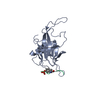
| |||||||||
|---|---|---|---|---|---|---|---|---|---|---|
| 1 |
| |||||||||
| NMR ensembles |
|
-Components
| #1: Protein | Mass: 18148.758 Da / Num. of mol.: 1 / Fragment: C-terminal FHA domain (FHA2) Source method: isolated from a genetically manipulated source Source: (gene. exp.) Gene: SPK1 or RAD53 / Plasmid: pGEX-4T / Species (production host): Escherichia coli / Production host:  References: UniProt: P22216, Transferases; Transferring phosphorus-containing groups; Phosphotransferases with an alcohol group as acceptor |
|---|---|
| #2: Protein/peptide | Mass: 1009.947 Da / Num. of mol.: 1 / Fragment: residues 826-832 / Source method: obtained synthetically Details: This phosphotyrosyl peptide was chemically synthesized. References: UniProt: P14737 |
| Has protein modification | Y |
-Experimental details
-Experiment
| Experiment | Method: SOLUTION NMR | ||||||||||||
|---|---|---|---|---|---|---|---|---|---|---|---|---|---|
| NMR experiment |
| ||||||||||||
| NMR details | Text: The structure was determined using triple-resonance NMR spectroscopy. |
- Sample preparation
| Details | Contents: 0.5 mM FHA2 U-15N,13C; 1mM phosphotyrosyl peptide of Rad9; 10 mM sodium phosphate (pH 6.5), 1 mM DTT, and 1 mM EDTA Solvent system: 95% H2O/5% D2O |
|---|---|
| Sample conditions | Ionic strength: 10 mM sodium phosphate(pH 6.5), 1 mM DTT, and 1 mM EDTA pH: 6.5 / Pressure: ambient / Temperature: 293 K |
-NMR measurement
| Radiation | Protocol: SINGLE WAVELENGTH / Monochromatic (M) / Laue (L): M |
|---|---|
| Radiation wavelength | Relative weight: 1 |
| NMR spectrometer | Type: Bruker DRX / Manufacturer: Bruker / Model: DRX / Field strength: 800 MHz |
- Processing
| NMR software |
| ||||||||||||||||||||
|---|---|---|---|---|---|---|---|---|---|---|---|---|---|---|---|---|---|---|---|---|---|
| Refinement | Method: simulated annealing / Software ordinal: 1 Details: The complex structures are generated using a total of 3398 restraints, 3210 distance restraints, and 188 TOLOS-derived dihedral angle restraints. | ||||||||||||||||||||
| NMR ensemble | Conformer selection criteria: structures with the lowest energy Conformers calculated total number: 100 / Conformers submitted total number: 22 |
 X-PLOR