- PDB-1qu5: NMR STRUCTURE OF A NEW PHOSPHOTYROSINE BINDING DOMAIN CONTAINING ... -
+
Open data
ID or keywords:
Loading...
-
Basic information
Entry
Database: PDB / ID: 1qu5
Title
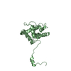
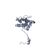
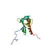
NMR STRUCTURE OF A NEW PHOSPHOTYROSINE BINDING DOMAIN CONTAINING THE FHA2 DOMAIN OF RAD 53
Components
PROTEIN KINASE SPK1
Keywords
TRANSFERASE / FHA / RAD53
Function / homology
Function and homology information
deoxyribonucleoside triphosphate biosynthetic process / meiotic recombination checkpoint signaling / dual-specificity kinase / telomere maintenance in response to DNA damage / negative regulation of DNA damage checkpoint / DNA replication origin binding / DNA replication initiation / regulation of DNA repair / protein serine/threonine/tyrosine kinase activity / DNA damage checkpoint signaling ...deoxyribonucleoside triphosphate biosynthetic process / meiotic recombination checkpoint signaling / dual-specificity kinase / telomere maintenance in response to DNA damage / negative regulation of DNA damage checkpoint / DNA replication origin binding / DNA replication initiation / regulation of DNA repair / protein serine/threonine/tyrosine kinase activity / DNA damage checkpoint signaling / intracellular protein localization / protein tyrosine kinase activity / protein kinase activity / protein serine kinase activity / DNA repair / protein serine/threonine kinase activity / ATP binding / nucleus / cytoplasm / cytosol Similarity search - Function
Serine/threonine-protein kinase Rad53 / Tumour Suppressor Smad4 - #20 / Tumour Suppressor Smad4 / Aurora kinase / Forkhead associated domain / Forkhead-associated (FHA) domain profile. / FHA domain / Forkhead-associated (FHA) domain / SMAD/FHA domain superfamily / Serine/threonine-protein kinase, active site ...Serine/threonine-protein kinase Rad53 / Tumour Suppressor Smad4 - #20 / Tumour Suppressor Smad4 / Aurora kinase / Forkhead associated domain / Forkhead-associated (FHA) domain profile. / FHA domain / Forkhead-associated (FHA) domain / SMAD/FHA domain superfamily / Serine/threonine-protein kinase, active site / Serine/Threonine protein kinases active-site signature. / Protein kinase domain / Serine/Threonine protein kinases, catalytic domain / Protein kinase, ATP binding site / Protein kinases ATP-binding region signature. / Protein kinase domain profile. / Protein kinase domain / Protein kinase-like domain superfamily / Sandwich / Mainly Beta Similarity search - Domain/homology
Method: SIMULATED ANNEALING, MOLECULAR DYNAMICS / Software ordinal: 1 Details: IN THE FIRST STAGE, THE SIMULATED ANNEALING STRUCTURES WERE DETERMINED BASED ON THE EXPERIMENTAL INTER PROTON DISTANCE RESTRAINTS (2651 IN TOTAL). THE RESULTING STRUCTURES WERE THEN USED AS ...Details: IN THE FIRST STAGE, THE SIMULATED ANNEALING STRUCTURES WERE DETERMINED BASED ON THE EXPERIMENTAL INTER PROTON DISTANCE RESTRAINTS (2651 IN TOTAL). THE RESULTING STRUCTURES WERE THEN USED AS INITIAL STRCUTURES FOR THE SECOND STAGE OF SIMULATED ANNEALING CALCULATIONS WHERE, IN ADDITION TO THE DISTANCE RESTRAINTS, THE STRUCTURES WERE REFINED AGAINST SECONDARY 13C(ALPHA)/13C(BETA) CHEMICAL SHIFT RESTRAINTS.
NMR representative
Selection criteria: minimized average structure
NMR ensemble
Conformer selection criteria: structures with the lowest energy Conformers calculated total number: 32 / Conformers submitted total number: 16
+
About Yorodumi
-
News
-
Feb 9, 2022. New format data for meta-information of EMDB entries
New format data for meta-information of EMDB entries
Version 3 of the EMDB header file is now the official format.
The previous official version 1.9 will be removed from the archive.
In the structure databanks used in Yorodumi, some data are registered as the other names, "COVID-19 virus" and "2019-nCoV". Here are the details of the virus and the list of structure data.
Jan 31, 2019. EMDB accession codes are about to change! (news from PDBe EMDB page)
EMDB accession codes are about to change! (news from PDBe EMDB page)
The allocation of 4 digits for EMDB accession codes will soon come to an end. Whilst these codes will remain in use, new EMDB accession codes will include an additional digit and will expand incrementally as the available range of codes is exhausted. The current 4-digit format prefixed with “EMD-” (i.e. EMD-XXXX) will advance to a 5-digit format (i.e. EMD-XXXXX), and so on. It is currently estimated that the 4-digit codes will be depleted around Spring 2019, at which point the 5-digit format will come into force.
The EM Navigator/Yorodumi systems omit the EMD- prefix.
Related info.:Q: What is EMD? / ID/Accession-code notation in Yorodumi/EM Navigator
Yorodumi is a browser for structure data from EMDB, PDB, SASBDB, etc.
This page is also the successor to EM Navigator detail page, and also detail information page/front-end page for Omokage search.
The word "yorodu" (or yorozu) is an old Japanese word meaning "ten thousand". "mi" (miru) is to see.
Related info.:EMDB / PDB / SASBDB / Comparison of 3 databanks / Yorodumi Search / Aug 31, 2016. New EM Navigator & Yorodumi / Yorodumi Papers / Jmol/JSmol / Function and homology information / Changes in new EM Navigator and Yorodumi
 Movie
Movie Controller

 Yorodumi
 Open data
 Basic information
 Components
 Keywords
 Function and homology information
 Authors
 Citation
 Structure visualization
 Downloads & links
 Other downloads
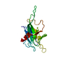


 PDBj
PDBj

 Assembly

 Sample preparation
 Processing
 X-PLOR