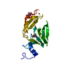
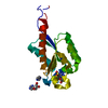
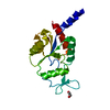
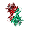
Entry Database : PDB / ID : 5zohTitle Crystal structure of a far-red light-absorbing form of AnPixJg2_BV4 in complex with biliverdin Methyl-accepting chemotaxis protein Keywords / / / / / Function / homology Function Domain/homology Component
/ / / / / / / / / / / / / / / / / / / / / / / / / / Biological species Nostoc sp. (bacteria)Method / / / Resolution : 1.6 Å Authors Miyazaki, T. / Fushimi, K. / Narikawa, R. Funding support Organization Grant number Country Japan Science and Technology
Journal : Proc. Natl. Acad. Sci. U.S.A. / Year : 2019Title : Rational conversion of chromophore selectivity of cyanobacteriochromes to accept mammalian intrinsic biliverdin.Authors : Fushimi, K. / Miyazaki, T. / Kuwasaki, Y. / Nakajima, T. / Yamamoto, T. / Suzuki, K. / Ueda, Y. / Miyake, K. / Takeda, Y. / Choi, J.H. / Kawagishi, H. / Park, E.Y. / Ikeuchi, M. / Sato, M. / Narikawa, R. History Deposition Apr 13, 2018 Deposition site / Processing site Revision 1.0 Apr 17, 2019 Provider / Type Revision 1.1 May 8, 2019 Group / Database references / Category / citation_authorItem _citation.journal_volume / _citation.page_first ... _citation.journal_volume / _citation.page_first / _citation.page_last / _citation_author.identifier_ORCID Revision 1.2 Nov 22, 2023 Group / Database references / Refinement descriptionCategory chem_comp_atom / chem_comp_bond ... chem_comp_atom / chem_comp_bond / database_2 / pdbx_initial_refinement_model Item / _database_2.pdbx_database_accessionRevision 1.3 Oct 30, 2024 Group / Category / pdbx_modification_feature
Show all Show less
 Movie
Movie Controller

 Yorodumi
 Open data
 Basic information
 Components
 Keywords
 Function and homology information
 Nostoc sp. (bacteria)
 X-RAY DIFFRACTION /
  Authors
 Japan, 1items
  Citation
 Structure visualization
 Downloads & links
 Other downloads
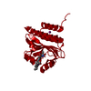

 PDBj
PDBj


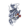
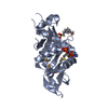
 Assembly
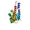

 Mass: 582.646 Da / Num. of mol.: 1 / Source method: obtained synthetically / Formula: C33H34N4O6
 Mass: 92.094 Da / Num. of mol.: 1 / Source method: obtained synthetically / Formula: C3H8O3
 Mass: 18.015 Da / Num. of mol.: 89 / Source method: isolated from a natural source / Formula: H2O
 Sample preparation
 Processing