 Movie
Movie Controller

[English] 日本語
 Yorodumi
- PDB-1izm: Structure of ygfB from Haemophilus influenzae (HI0817), a Conserv... -
+ Open data

- Basic information
| Entry | Database: PDB / ID: 1izm | ||||||
|---|---|---|---|---|---|---|---|
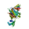
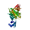
| Title | Structure of ygfB from Haemophilus influenzae (HI0817), a Conserved Hypothetical Protein | ||||||
 Components Components | HYPOTHETICAL PROTEIN HI0817 | ||||||
 Keywords Keywords | STRUCTURAL GENOMICS / UNKNOWN FUNCTION / hypothetical protein / HI0817 / ygfB / Structure 2 Function Project / S2F | ||||||
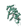
| Function / homology | YgfB uncharacterised protein family PF03695 / YgfB-like / YgfB-like superfamily / Uncharacterised protein family (UPF0149) / Four Helix Bundle (Hemerythrin (Met), subunit A) / Up-down Bundle / Mainly Alpha / cytosol / UPF0149 protein HI_0817 Function and homology information Function and homology information | ||||||
| Biological species |  Haemophilus influenzae (bacteria) Haemophilus influenzae (bacteria) | ||||||
| Method |  X-RAY DIFFRACTION / SYNCHROTRON / MAD / Resolution: 1.95 Å X-RAY DIFFRACTION / SYNCHROTRON / MAD / Resolution: 1.95 Å | ||||||
 Authors Authors | Galkin, A. / Sarikaya, E. / Lehmann, C. / Howard, A. / Herzberg, O. / Structure 2 Function Project (S2F) | ||||||
 Citation Citation | Journal: Proteins / Year: 2004 Title: X-ray structure of HI0817 from Haemophilus influenzae: protein of unknown function with a novel fold. Authors: Galkin, A. / Sarikaya, E. / Lehmann, C. / Howard, A. / Herzberg, O. | ||||||
| History |
|
- Structure visualization
| Structure viewer | Molecule: MolmilJmol/JSmol |
|---|
- Downloads & links
-Download
| PDBx/mmCIF format | 1izm.cif.gz | 51.4 KB | Display | PDBx/mmCIF format |
|---|---|---|---|---|
| PDB format | pdb1izm.ent.gz | 36.5 KB | Display | PDB format |
| PDBx/mmJSON format | 1izm.json.gz | Tree view | PDBx/mmJSON format | |
| Others |  Other downloads Other downloads |
-Validation report
| Arichive directory | https://data.pdbj.org/pub/pdb/validation_reports/iz/1izmftp://data.pdbj.org/pub/pdb/validation_reports/iz/1izm | HTTPS FTP |
|---|
-Related structure data
| Similar structure data | |
|---|---|
| Other databases |


-Links
 PDBj
PDBj- Assembly
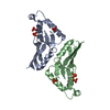
| Deposited unit | 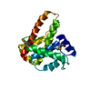
| ||||||||
|---|---|---|---|---|---|---|---|---|---|
| 1 | 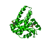
| ||||||||
| Unit cell |
| ||||||||
| Details | Dimer.The asymmetric unit contains one monomer. The dimer is generated from the monomer by a crystallographic symmetry: Y-1, X+1,-Z+1 |
-Components
| #1: Protein | Mass: 20604.152 Da / Num. of mol.: 1 / Mutation: L8M Source method: isolated from a genetically manipulated source Source: (gene. exp.) Haemophilus influenzae (bacteria) / Gene: HI0817 / Plasmid: pET100 / Production host: |
|---|---|
| #2: Water | ChemComp-HOH /  Mass: 18.015 Da / Num. of mol.: 201 / Source method: isolated from a natural source / Formula: H2O Mass: 18.015 Da / Num. of mol.: 201 / Source method: isolated from a natural source / Formula: H2O |
| Has protein modification | Y |
-Experimental details
-Experiment
| Experiment | Method: X-RAY DIFFRACTION / Number of used crystals: 1 |
|---|
- Sample preparation
| Crystal | Density Matthews: 2.15 Å3/Da / Density % sol: 42.92 % | ||||||||||||||||||||||||||||||||||||||||||
|---|---|---|---|---|---|---|---|---|---|---|---|---|---|---|---|---|---|---|---|---|---|---|---|---|---|---|---|---|---|---|---|---|---|---|---|---|---|---|---|---|---|---|---|
| Crystal grow | Temperature: 277 K / Method: vapor diffusion, hanging drop / pH: 4.8 Details: 1.4 M ammonimum sulfate, 0.05 M potassium phosphate, 3% iso-propanol, pH 4.8, VAPOR DIFFUSION, HANGING DROP, temperature 277K | ||||||||||||||||||||||||||||||||||||||||||
| Crystal grow | *PLUS Temperature: 4 ℃ / pH: 7 / Method: vapor diffusion, hanging drop | ||||||||||||||||||||||||||||||||||||||||||
| Components of the solutions | *PLUS
|
-Data collection
| Diffraction | Mean temperature: 100 K | ||||||||||||
|---|---|---|---|---|---|---|---|---|---|---|---|---|---|
| Diffraction source | Source: SYNCHROTRON / Site: APS  / Beamline: 17-ID / Wavelength: 0.97946, 0.97961, 0.96863 / Beamline: 17-ID / Wavelength: 0.97946, 0.97961, 0.96863 | ||||||||||||
| Detector | Type: ADSC QUANTUM 4 / Detector: CCD / Date: Dec 6, 2001 | ||||||||||||
| Radiation | Monochromator: Si(111) double crystal system / Protocol: MAD / Monochromatic (M) / Laue (L): M / Scattering type: x-ray | ||||||||||||
| Radiation wavelength |
| ||||||||||||
| Reflection | Resolution: 1.95→20 Å / Num. all: 14167 / Num. obs: 14167 / % possible obs: 97.9 % / Observed criterion σ(F): 0 / Observed criterion σ(I): 0 / Rmerge(I) obs: 0.05 / Net I/σ(I): 21.1 | ||||||||||||
| Reflection shell | Resolution: 1.95→2.02 Å / Rmerge(I) obs: 0.097 / % possible all: 91.9 | ||||||||||||
| Reflection | *PLUS Num. obs: 24638 / % possible obs: 98 % / Num. measured all: 298675 / Rmerge(I) obs: 0.05 | ||||||||||||
| Reflection shell | *PLUS % possible obs: 92 % |
- Processing
| Software |
| |||||||||||||||||||||||||
|---|---|---|---|---|---|---|---|---|---|---|---|---|---|---|---|---|---|---|---|---|---|---|---|---|---|---|
| Refinement | Method to determine structure: MAD / Resolution: 1.95→20 Å / Cross valid method: THROUGHOUT / σ(F): 0 / Stereochemistry target values: Engh & Huber
| |||||||||||||||||||||||||
| Displacement parameters | Biso mean: 38 Å2 | |||||||||||||||||||||||||
| Refinement step | Cycle: LAST / Resolution: 1.95→20 Å
| |||||||||||||||||||||||||
| Refine LS restraints |
| |||||||||||||||||||||||||
| LS refinement shell | Resolution: 1.95→2.02 Å
| |||||||||||||||||||||||||
| Refine LS restraints | *PLUS Type: c_bond_d / Dev ideal: 0.02 |
