 Movie
Movie Controller

[English] 日本語
 Yorodumi
- PDB-3v7r: Crystal structure of Staphylococcus aureus biotin protein ligase ... -
+ Open data

- Basic information
| Entry | Database: PDB / ID: 3v7r | ||||||
|---|---|---|---|---|---|---|---|
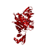
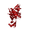
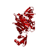
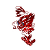
| Title | Crystal structure of Staphylococcus aureus biotin protein ligase in complex with inhibitor | ||||||
 Components Components | Biotin ligase | ||||||
 Keywords Keywords | LIGASE/LIGASE INHIBITOR / biotin / metabolism / biotin carboxyl carrier protein / LIGASE-LIGASE INHIBITOR complex | ||||||
| Function / homology |  Function and homology information Function and homology informationSH3 type barrels. - #100 / Bira Bifunctional Protein; Domain 2 / BirA Bifunctional Protein; domain 2 / Winged helix-like DNA-binding domain superfamily/Winged helix DNA-binding domain / SH3 type barrels. / Arc Repressor Mutant, subunit A / Roll / 2-Layer Sandwich / Orthogonal Bundle / Mainly Beta ...SH3 type barrels. - #100 / Bira Bifunctional Protein; Domain 2 / BirA Bifunctional Protein; domain 2 / Winged helix-like DNA-binding domain superfamily/Winged helix DNA-binding domain / SH3 type barrels. / Arc Repressor Mutant, subunit A / Roll / 2-Layer Sandwich / Orthogonal Bundle / Mainly Beta / Mainly Alpha / Alpha Beta Similarity search - Domain/homology | ||||||
| Biological species |   Staphylococcus aureus (bacteria) Staphylococcus aureus (bacteria) | ||||||
| Method |  X-RAY DIFFRACTION / SYNCHROTRON / MOLECULAR REPLACEMENT / Resolution: 2.61 Å X-RAY DIFFRACTION / SYNCHROTRON / MOLECULAR REPLACEMENT / Resolution: 2.61 Å | ||||||
 Authors Authors | Yap, M.Y. / Pendini, N.R. | ||||||
 Citation Citation | Journal: J.Biol.Chem. / Year: 2012 Title: Selective inhibition of biotin protein ligase from Staphylococcus aureus. Authors: Soares da Costa, T.P. / Tieu, W. / Yap, M.Y. / Pendini, N.R. / Polyak, S.W. / Sejer Pedersen, D. / Morona, R. / Turnidge, J.D. / Wallace, J.C. / Wilce, M.C. / Booker, G.W. / Abell, A.D. | ||||||
| History |
|
- Structure visualization
| Structure viewer | Molecule: MolmilJmol/JSmol |
|---|
- Downloads & links
-Download
| PDBx/mmCIF format | 3v7r.cif.gz | 83.7 KB | Display | PDBx/mmCIF format |
|---|---|---|---|---|
| PDB format | pdb3v7r.ent.gz | 61.4 KB | Display | PDB format |
| PDBx/mmJSON format | 3v7r.json.gz | Tree view | PDBx/mmJSON format | |
| Others |  Other downloads Other downloads |
-Validation report
| Arichive directory | https://data.pdbj.org/pub/pdb/validation_reports/v7/3v7rftp://data.pdbj.org/pub/pdb/validation_reports/v7/3v7r | HTTPS FTP |
|---|
-Related structure data
| Related structure data |  3v7cC  3v7sC  3v8jC  3v8kC  3v8lC  4dq2C  3rkwS C: citing same article ( S: Starting model for refinement |
|---|---|
| Similar structure data |


-Links
 PDBj
PDBj- Assembly
| Deposited unit | 
| ||||||||
|---|---|---|---|---|---|---|---|---|---|
| 1 | 
| ||||||||
| Unit cell |
| ||||||||
| Components on special symmetry positions |
|
-Components
| #1: Protein | Mass: 38014.836 Da / Num. of mol.: 1 Source method: isolated from a genetically manipulated source Source: (gene. exp.) Staphylococcus aureus (bacteria) / Strain: A9781 / Gene: SAOG_00031 / Production host: |
|---|---|
| #2: Chemical | ChemComp-32G / (  Mass: 470.594 Da / Num. of mol.: 1 / Source method: obtained synthetically / Formula: C21H30N10OS Mass: 470.594 Da / Num. of mol.: 1 / Source method: obtained synthetically / Formula: C21H30N10OS |
| #3: Water | ChemComp-HOH /  Mass: 18.015 Da / Num. of mol.: 146 / Source method: isolated from a natural source / Formula: H2O Mass: 18.015 Da / Num. of mol.: 146 / Source method: isolated from a natural source / Formula: H2O |
-Experimental details
-Experiment
| Experiment | Method: X-RAY DIFFRACTION / Number of used crystals: 1 |
|---|
- Sample preparation
| Crystal | Density Matthews: 3.85 Å3/Da / Density % sol: 68.01 % |
|---|---|
| Crystal grow | Temperature: 277 K / Method: vapor diffusion, hanging drop / pH: 7.5 Details: 10% PEG 8000, 10% glycerol, pH 7.5, VAPOR DIFFUSION, HANGING DROP, temperature 277K |
-Data collection
| Diffraction | Mean temperature: 277 K |
|---|---|
| Diffraction source | Source: SYNCHROTRON / Site: Australian Synchrotron  / Beamline: MX1 / Wavelength: 1.54 Å / Beamline: MX1 / Wavelength: 1.54 Å |
| Detector | Date: Sep 21, 2009 |
| Radiation | Protocol: SINGLE WAVELENGTH / Monochromatic (M) / Laue (L): M / Scattering type: x-ray |
| Radiation wavelength | Wavelength: 1.54 Å / Relative weight: 1 |
| Reflection | Resolution: 2.61→19.85 Å / Num. obs: 18442 |
- Processing
| Software | Name: REFMAC / Version: 5.5.0102 / Classification: refinement | ||||||||||||||||||||||||||||||||||||||||||||||||||||||||||||||||||||||||||||||||||||||||||||||||||||||||||||||||||||||||||||||||||||||||||||||||||||||||||||||||||||||||||
|---|---|---|---|---|---|---|---|---|---|---|---|---|---|---|---|---|---|---|---|---|---|---|---|---|---|---|---|---|---|---|---|---|---|---|---|---|---|---|---|---|---|---|---|---|---|---|---|---|---|---|---|---|---|---|---|---|---|---|---|---|---|---|---|---|---|---|---|---|---|---|---|---|---|---|---|---|---|---|---|---|---|---|---|---|---|---|---|---|---|---|---|---|---|---|---|---|---|---|---|---|---|---|---|---|---|---|---|---|---|---|---|---|---|---|---|---|---|---|---|---|---|---|---|---|---|---|---|---|---|---|---|---|---|---|---|---|---|---|---|---|---|---|---|---|---|---|---|---|---|---|---|---|---|---|---|---|---|---|---|---|---|---|---|---|---|---|---|---|---|---|---|
| Refinement | Method to determine structure: MOLECULAR REPLACEMENT Starting model: 3RKW Resolution: 2.61→19.85 Å / Cor.coef. Fo:Fc: 0.932 / Cor.coef. Fo:Fc free: 0.899 / SU B: 8.838 / SU ML: 0.189 / Cross valid method: THROUGHOUT / ESU R: 0.353 / ESU R Free: 0.266 / Stereochemistry target values: MAXIMUM LIKELIHOOD / Details: HYDROGENS HAVE BEEN ADDED IN THE RIDING POSITIONS
| ||||||||||||||||||||||||||||||||||||||||||||||||||||||||||||||||||||||||||||||||||||||||||||||||||||||||||||||||||||||||||||||||||||||||||||||||||||||||||||||||||||||||||
| Solvent computation | Ion probe radii: 0.8 Å / Shrinkage radii: 0.8 Å / VDW probe radii: 1.4 Å / Solvent model: MASK | ||||||||||||||||||||||||||||||||||||||||||||||||||||||||||||||||||||||||||||||||||||||||||||||||||||||||||||||||||||||||||||||||||||||||||||||||||||||||||||||||||||||||||
| Displacement parameters | Biso mean: 52.426 Å2
| ||||||||||||||||||||||||||||||||||||||||||||||||||||||||||||||||||||||||||||||||||||||||||||||||||||||||||||||||||||||||||||||||||||||||||||||||||||||||||||||||||||||||||
| Refinement step | Cycle: LAST / Resolution: 2.61→19.85 Å
| ||||||||||||||||||||||||||||||||||||||||||||||||||||||||||||||||||||||||||||||||||||||||||||||||||||||||||||||||||||||||||||||||||||||||||||||||||||||||||||||||||||||||||
| Refine LS restraints |
| ||||||||||||||||||||||||||||||||||||||||||||||||||||||||||||||||||||||||||||||||||||||||||||||||||||||||||||||||||||||||||||||||||||||||||||||||||||||||||||||||||||||||||
| LS refinement shell | Resolution: 2.612→2.679 Å / Total num. of bins used: 20
|
