 Movie
Movie Controller


+ Open data

- Basic information
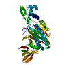
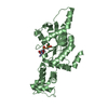
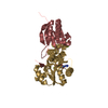
| Entry | Database: PDB / ID: 1hxd | ||||||
|---|---|---|---|---|---|---|---|
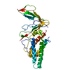
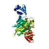
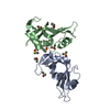
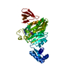
| Title | CRYSTAL STRUCTURE OF E. COLI BIOTIN REPRESSOR WITH BOUND BIOTIN | ||||||
 Components Components | BIRA BIFUNCTIONAL PROTEIN | ||||||
 Keywords Keywords | LIGASE / Repressor / BIOTIN / DNA-Binding | ||||||
| Function / homology |  Function and homology information Function and homology informationbiotin metabolic process / biotin-[biotin carboxyl-carrier protein] ligase / biotin--[biotin carboxyl-carrier protein] ligase activity / biotin biosynthetic process / biotin binding / transcription repressor complex / nucleic acid binding / transcription cis-regulatory region binding / regulation of DNA-templated transcription / protein homodimerization activity ...biotin metabolic process / biotin-[biotin carboxyl-carrier protein] ligase / biotin--[biotin carboxyl-carrier protein] ligase activity / biotin biosynthetic process / biotin binding / transcription repressor complex / nucleic acid binding / transcription cis-regulatory region binding / regulation of DNA-templated transcription / protein homodimerization activity / DNA binding / ATP binding / cytoplasm Similarity search - Function | ||||||
| Biological species |  | ||||||
| Method |  X-RAY DIFFRACTION / MOLECULAR REPLACEMENT / Resolution: 2.4 Å X-RAY DIFFRACTION / MOLECULAR REPLACEMENT / Resolution: 2.4 Å | ||||||
 Authors Authors | Kwon, K. / Streaker, E.D. / Ruparelia, S. / Beckett, D. | ||||||
 Citation Citation | Journal: Proc.Natl.Acad.Sci.USA / Year: 2001 Title: Corepressor-induced organization and assembly of the biotin repressor: a model for allosteric activation of a transcriptional regulator. Authors: Weaver, L.H. / Kwon, K. / Beckett, D. / Matthews, B.W. #1: Journal: J.Mol.Biol. / Year: 2000Title: MULTIPLE DISORDERED LOOPS FUNCTION IN COREPRESOR-INDUCED DIMERIZATION OF THE BIOTIN REPRESSOR Authors: Kwon, K. / Streaker, E.D. / Ruparelia, S. / Beckett, D. #2: Journal: Proc.Natl.Acad.Sci.USA / Year: 1992Title: E. COLI BIOTIN HOLOENZYME SYNTHETASE/BIO REPRESSOR CRYSTAL STRUCTURE DELINEATES THE BIOTIN- AND DNA-BINDING DOMAINS Authors: Wilson, K.P. / Shewchuk, L.M. / Brennan, R.G. / Otsuka, A.J. / Matthews, B.W. | ||||||
| History |
|
- Structure visualization
| Structure viewer | Molecule: MolmilJmol/JSmol |
|---|
- Downloads & links
-Download
| PDBx/mmCIF format | 1hxd.cif.gz | 131.3 KB | Display | PDBx/mmCIF format |
|---|---|---|---|---|
| PDB format | pdb1hxd.ent.gz | 102.5 KB | Display | PDB format |
| PDBx/mmJSON format | 1hxd.json.gz | Tree view | PDBx/mmJSON format | |
| Others |  Other downloads Other downloads |
-Validation report
| Arichive directory | https://data.pdbj.org/pub/pdb/validation_reports/hx/1hxdftp://data.pdbj.org/pub/pdb/validation_reports/hx/1hxd | HTTPS FTP |
|---|
-Related structure data
| Related structure data | 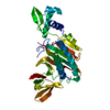 1biaS S: Starting model for refinement |
|---|---|
| Similar structure data |


-Links
 PDBj
PDBj
- Assembly
| Deposited unit | 
| ||||||||
|---|---|---|---|---|---|---|---|---|---|
| 1 | 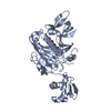
| ||||||||
| 2 | 
| ||||||||
| Unit cell |
|
-Components
| #1: Protein | Mass: 35351.918 Da / Num. of mol.: 2 Source method: isolated from a genetically manipulated source Source: (gene. exp.) References: UniProt: P06709, biotin-[biotin carboxyl-carrier protein] ligase #2: Chemical |   Mass: 244.311 Da / Num. of mol.: 2 / Source method: obtained synthetically / Formula: C10H16N2O3S Mass: 244.311 Da / Num. of mol.: 2 / Source method: obtained synthetically / Formula: C10H16N2O3S#3: Water | ChemComp-HOH / |  Mass: 18.015 Da / Num. of mol.: 59 / Source method: isolated from a natural source / Formula: H2O Mass: 18.015 Da / Num. of mol.: 59 / Source method: isolated from a natural source / Formula: H2O |
|---|
-Experimental details
-Experiment
| Experiment | Method: X-RAY DIFFRACTION / Number of used crystals: 1 |
|---|
- Sample preparation
| Crystal | Density Matthews: 2.89 Å3/Da / Density % sol: 57.46 % | |||||||||||||||
|---|---|---|---|---|---|---|---|---|---|---|---|---|---|---|---|---|
| Crystal grow | *PLUS Temperature: 4 ℃ / pH: 6.5 / Method: vapor diffusion, hanging drop | |||||||||||||||
| Components of the solutions | *PLUS
|
-Data collection
| Diffraction source | Source: ROTATING ANODE / Type: RIGAKU RU200 / Wavelength: 1.5418 Å |
|---|---|
| Detector | Type: UCSD MARK II / Detector: AREA DETECTOR / Date: Sep 2, 1999 |
| Radiation | Monochromator: GRAPHITE / Protocol: SINGLE WAVELENGTH / Monochromatic (M) / Laue (L): M / Scattering type: x-ray |
| Radiation wavelength | Wavelength: 1.5418 Å / Relative weight: 1 |
| Reflection | Resolution: 2.4→13 Å / Num. all: 48934 / Num. obs: 26762 / % possible obs: 83 % / Rmerge(I) obs: 0.071 / Net I/σ(I): 9 |
| Reflection shell | Resolution: 2.4→2.59 Å / Rmerge(I) obs: 0.183 / Mean I/σ(I) obs: 1.8 / Num. unique all: 4098 |
| Reflection | *PLUS % possible obs: 83 % / Num. measured all: 48934 |
| Reflection shell | *PLUS % possible obs: 33 % |
- Processing
| Software |
| ||||||||||||||||
|---|---|---|---|---|---|---|---|---|---|---|---|---|---|---|---|---|---|
| Refinement | Method to determine structure: MOLECULAR REPLACEMENT Starting model: PDB entry 1BIA Resolution: 2.4→13 Å / σ(F): 0
| ||||||||||||||||
| Refinement step | Cycle: LAST / Resolution: 2.4→13 Å
| ||||||||||||||||
| Refine LS restraints |
| ||||||||||||||||
| Software | *PLUS Name: TNT / Classification: refinement | ||||||||||||||||
| Refinement | *PLUS Highest resolution: 2.4 Å / Lowest resolution: 13 Å / σ(F): 0 / Rfactor all: 0.189 | ||||||||||||||||
| Solvent computation | *PLUS | ||||||||||||||||
| Displacement parameters | *PLUS | ||||||||||||||||
| Refine LS restraints | *PLUS Type: t_angle_deg / Dev ideal: 2.8 |
