 Movie
Movie Controller

[English] 日本語
 Yorodumi
- PDB-1gpp: Crystal structure of the S.cerevisiae Homing Endonuclease PI-SceI... -
+ Open data

- Basic information
| Entry | Database: PDB / ID: 1gpp | ||||||
|---|---|---|---|---|---|---|---|
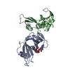
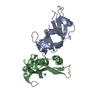
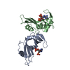
| Title | Crystal structure of the S.cerevisiae Homing Endonuclease PI-SceI Domain I | ||||||
 Components Components | ENDONUCLEASE PI-SCEI | ||||||
 Keywords Keywords | ENDONUCLEASE / HOMING / PROTEIN SPLICING | ||||||
| Function / homology |  Function and homology information Function and homology informationInsulin receptor recycling / Transferrin endocytosis and recycling / ROS and RNS production in phagocytes / Golgi lumen acidification / Amino acids regulate mTORC1 / vacuolar proton-transporting V-type ATPase, V1 domain / endosomal lumen acidification / proton-transporting V-type ATPase complex / intron homing / vacuolar proton-transporting V-type ATPase complex ...Insulin receptor recycling / Transferrin endocytosis and recycling / ROS and RNS production in phagocytes / Golgi lumen acidification / Amino acids regulate mTORC1 / vacuolar proton-transporting V-type ATPase, V1 domain / endosomal lumen acidification / proton-transporting V-type ATPase complex / intron homing / vacuolar proton-transporting V-type ATPase complex / intein-mediated protein splicing / vacuolar acidification / fungal-type vacuole membrane / proton-transporting ATPase activity, rotational mechanism / H+-transporting two-sector ATPase / ATP metabolic process / proton transmembrane transport / endonuclease activity / Hydrolases; Acting on ester bonds / Golgi membrane / hydrolase activity / mRNA binding / DNA binding / ATP binding Similarity search - Function | ||||||
| Biological species |  | ||||||
| Method |  X-RAY DIFFRACTION / SYNCHROTRON / MOLECULAR REPLACEMENT / Resolution: 1.35 Å X-RAY DIFFRACTION / SYNCHROTRON / MOLECULAR REPLACEMENT / Resolution: 1.35 Å | ||||||
 Authors Authors | Werner, E. / Wende, W. / Pingoud, A. / Heinemann, U. | ||||||
 Citation Citation | Journal: Nucleic Acids Res. / Year: 2002 Title: High Resolution Crystal Structure of Domain I of the Saccharomyces Cerevisiae Homing Endonuclease Pi-Scei Authors: Werner, E. / Wende, W. / Pingoud, A. / Heinemann, U. | ||||||
| History |
| ||||||
| Remark 700 | SHEET DETERMINATION METHOD: DSSP THE SHEETS PRESENTED AS "AA" IN EACH CHAIN ON SHEET RECORDS BELOW ... SHEET DETERMINATION METHOD: DSSP THE SHEETS PRESENTED AS "AA" IN EACH CHAIN ON SHEET RECORDS BELOW IS ACTUALLY AN 7-STRANDED BARREL THIS IS REPRESENTED BY A 8-STRANDED SHEET IN WHICH THE FIRST AND LAST STRANDS ARE IDENTICAL. |
- Structure visualization
| Structure viewer | Molecule: MolmilJmol/JSmol |
|---|
- Downloads & links
-Download
| PDBx/mmCIF format | 1gpp.cif.gz | 114.8 KB | Display | PDBx/mmCIF format |
|---|---|---|---|---|
| PDB format | pdb1gpp.ent.gz | 88.1 KB | Display | PDB format |
| PDBx/mmJSON format | 1gpp.json.gz | Tree view | PDBx/mmJSON format | |
| Others |  Other downloads Other downloads |
-Validation report
| Arichive directory | https://data.pdbj.org/pub/pdb/validation_reports/gp/1gppftp://data.pdbj.org/pub/pdb/validation_reports/gp/1gpp | HTTPS FTP |
|---|
-Related structure data
| Related structure data | 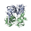 1vdeS S: Starting model for refinement |
|---|---|
| Similar structure data |


-Links
 PDBj
PDBj


- Assembly
| Deposited unit | 
| ||||||||
|---|---|---|---|---|---|---|---|---|---|
| 1 |
| ||||||||
| Unit cell |
|
-Components
| #1: Protein | Mass: 27034.643 Da / Num. of mol.: 1 Fragment: PROTEIN SPLICING DOMAIN, RESIDUES 284-466,693-736, SEE REMARK 999 Mutation: YES Source method: isolated from a genetically manipulated source Details: GLY183 LINKS ILE182 AND ALA410, WHERE IN THE FULL LENGTH PROTEIN IS DOMAIN II Source: (gene. exp.) Production host:  References: UniProt: P17255, H+-transporting two-sector ATPase |
|---|---|
| #2: Water | ChemComp-HOH /  Mass: 18.015 Da / Num. of mol.: 303 / Source method: isolated from a natural source / Formula: H2O Mass: 18.015 Da / Num. of mol.: 303 / Source method: isolated from a natural source / Formula: H2O |
| Compound details | MUTATIONS: ARG327SER, VAL350MET, ILE415VAL, LEU466GLY |
| Sequence details | THE PROTEIN PI-SCEI IS AN INTEIN OF THE PRIMARY TRANSLATION PRODUCT OF THE ATP SYNTHASE (SWS ENTRY ...THE PROTEIN PI-SCEI IS AN INTEIN OF THE PRIMARY TRANSLATIO |
-Experimental details
-Experiment
| Experiment | Method: X-RAY DIFFRACTION / Number of used crystals: 1 |
|---|
- Sample preparation
| Crystal | Density Matthews: 2.7 Å3/Da / Density % sol: 54 % | ||||||||||||||||||||||||||||||
|---|---|---|---|---|---|---|---|---|---|---|---|---|---|---|---|---|---|---|---|---|---|---|---|---|---|---|---|---|---|---|---|
| Crystal grow | pH: 4.8 Details: 30 % PEG4000, 0.1 M SODIUM CITRATE PH 5.6, 0.2 M NH4-ACETATE | ||||||||||||||||||||||||||||||
| Crystal grow | *PLUS pH: 5.6 / Method: vapor diffusion, hanging drop | ||||||||||||||||||||||||||||||
| Components of the solutions | *PLUS
|
-Data collection
| Diffraction | Mean temperature: 100 K |
|---|---|
| Diffraction source | Source: SYNCHROTRON / Site: EMBL/DESY, HAMBURG  / Beamline: BW7B / Wavelength: 0.8453 / Beamline: BW7B / Wavelength: 0.8453 |
| Detector | Type: MARRESEARCH / Detector: IMAGE PLATE / Date: May 15, 2001 / Details: MIRRORS |
| Radiation | Protocol: SINGLE WAVELENGTH / Monochromatic (M) / Laue (L): M / Scattering type: x-ray |
| Radiation wavelength | Wavelength: 0.8453 Å / Relative weight: 1 |
| Reflection | Resolution: 1.35→20 Å / Num. obs: 51602 / % possible obs: 94.1 % / Redundancy: 2.5 % / Rmerge(I) obs: 0.029 / Net I/σ(I): 34.5 |
| Reflection shell | Resolution: 1.35→1.37 Å / Redundancy: 1.5 % / Rmerge(I) obs: 0.164 / Mean I/σ(I) obs: 2.5 / % possible all: 64.1 |
| Reflection | *PLUS Lowest resolution: 20 Å / Num. measured all: 359618 |
| Reflection shell | *PLUS % possible obs: 64.1 % / Num. unique obs: 1141 |
- Processing
| Software |
| ||||||||||||||||||||||||||||||||||||||||||||||||||||||||||||||||||||||||||||||||||||||||||||||||||||||||||||||||||||||||||||||||||||||||||||||||||||||||||||||||||||||||||||||||||||||
|---|---|---|---|---|---|---|---|---|---|---|---|---|---|---|---|---|---|---|---|---|---|---|---|---|---|---|---|---|---|---|---|---|---|---|---|---|---|---|---|---|---|---|---|---|---|---|---|---|---|---|---|---|---|---|---|---|---|---|---|---|---|---|---|---|---|---|---|---|---|---|---|---|---|---|---|---|---|---|---|---|---|---|---|---|---|---|---|---|---|---|---|---|---|---|---|---|---|---|---|---|---|---|---|---|---|---|---|---|---|---|---|---|---|---|---|---|---|---|---|---|---|---|---|---|---|---|---|---|---|---|---|---|---|---|---|---|---|---|---|---|---|---|---|---|---|---|---|---|---|---|---|---|---|---|---|---|---|---|---|---|---|---|---|---|---|---|---|---|---|---|---|---|---|---|---|---|---|---|---|---|---|---|---|
| Refinement | Method to determine structure: MOLECULAR REPLACEMENT Starting model: PDB ENTRY 1VDE Resolution: 1.35→20 Å / Cor.coef. Fo:Fc: 0.976 / Cor.coef. Fo:Fc free: 0.963 / SU B: 2.249 / SU ML: 0.048 / Cross valid method: THROUGHOUT / ESU R: 0.056 / ESU R Free: 0.055 / Stereochemistry target values: MAXIMUM LIKELIHOOD Details: HYDROGENS HAVE BEEN ADDED IN THE RIDING POSITIONS. FIRST 8 RESIDUES OF THE HIS-TAG AND RESIDUES 55 - 66 ARE NOT VISIBLE IN ELECTRON DENSITY.
| ||||||||||||||||||||||||||||||||||||||||||||||||||||||||||||||||||||||||||||||||||||||||||||||||||||||||||||||||||||||||||||||||||||||||||||||||||||||||||||||||||||||||||||||||||||||
| Solvent computation | Ion probe radii: 0.8 Å / Shrinkage radii: 0.8 Å / VDW probe radii: 1.4 Å / Solvent model: BABINET MODEL WITH MASK | ||||||||||||||||||||||||||||||||||||||||||||||||||||||||||||||||||||||||||||||||||||||||||||||||||||||||||||||||||||||||||||||||||||||||||||||||||||||||||||||||||||||||||||||||||||||
| Displacement parameters | Biso mean: 19.93 Å2
| ||||||||||||||||||||||||||||||||||||||||||||||||||||||||||||||||||||||||||||||||||||||||||||||||||||||||||||||||||||||||||||||||||||||||||||||||||||||||||||||||||||||||||||||||||||||
| Refinement step | Cycle: LAST / Resolution: 1.35→20 Å
| ||||||||||||||||||||||||||||||||||||||||||||||||||||||||||||||||||||||||||||||||||||||||||||||||||||||||||||||||||||||||||||||||||||||||||||||||||||||||||||||||||||||||||||||||||||||
| Refine LS restraints |
|