 Movie
Movie Controller

[English] 日本語
 Yorodumi
- PDB-3rkx: Structural characterisation of staphylococcus aureus biotin prote... -
+ Open data

- Basic information
| Entry | Database: PDB / ID: 3rkx | ||||||
|---|---|---|---|---|---|---|---|
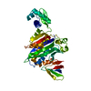
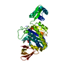
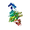
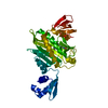
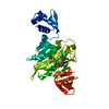
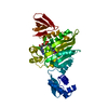
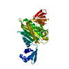
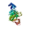
| Title | Structural characterisation of staphylococcus aureus biotin protein ligase | ||||||
 Components Components | Biotin-[acetyl-CoA-carboxylase] ligase | ||||||
 Keywords Keywords | LIGASE / biotin protein ligase / 3 domains / enzyme DNA binding / biotin carrier coupling domains | ||||||
| Function / homology |  Function and homology information Function and homology informationSH3 type barrels. - #100 / Bira Bifunctional Protein; Domain 2 / BirA Bifunctional Protein; domain 2 / Winged helix-like DNA-binding domain superfamily/Winged helix DNA-binding domain / SH3 type barrels. / Arc Repressor Mutant, subunit A / Roll / 2-Layer Sandwich / Orthogonal Bundle / Mainly Beta ...SH3 type barrels. - #100 / Bira Bifunctional Protein; Domain 2 / BirA Bifunctional Protein; domain 2 / Winged helix-like DNA-binding domain superfamily/Winged helix DNA-binding domain / SH3 type barrels. / Arc Repressor Mutant, subunit A / Roll / 2-Layer Sandwich / Orthogonal Bundle / Mainly Beta / Mainly Alpha / Alpha Beta Similarity search - Domain/homology | ||||||
| Biological species |   Staphylococcus aureus (bacteria) Staphylococcus aureus (bacteria) | ||||||
| Method |  X-RAY DIFFRACTION / SYNCHROTRON / MOLECULAR REPLACEMENT / Resolution: 2.1 Å X-RAY DIFFRACTION / SYNCHROTRON / MOLECULAR REPLACEMENT / Resolution: 2.1 Å | ||||||
 Authors Authors | Wilce, M.C.J. | ||||||
 Citation Citation | Journal: TO BE PUBLISHED Title: Structural characterisation of staphylococcus aureus biotin protein ligase Authors: Pendini, N.R. / Yap, M.Y. / Polyak, S.W. / Cowieson, N. / Traore, D.A.K. / Booker, G.W. / Wallace, J.C. / Abell, A. / Wilce, J.A. / Wilce, M.C.J. | ||||||
| History |
|
- Structure visualization
| Structure viewer | Molecule: MolmilJmol/JSmol |
|---|
- Downloads & links
-Download
| PDBx/mmCIF format | 3rkx.cif.gz | 138.5 KB | Display | PDBx/mmCIF format |
|---|---|---|---|---|
| PDB format | pdb3rkx.ent.gz | 107.4 KB | Display | PDB format |
| PDBx/mmJSON format | 3rkx.json.gz | Tree view | PDBx/mmJSON format | |
| Others |  Other downloads Other downloads |
-Validation report
| Arichive directory | https://data.pdbj.org/pub/pdb/validation_reports/rk/3rkxftp://data.pdbj.org/pub/pdb/validation_reports/rk/3rkx | HTTPS FTP |
|---|
-Related structure data
| Related structure data |  3rkwC  3rkyC  3rirS S: Starting model for refinement C: citing same article ( |
|---|---|
| Similar structure data |


-Links
 PDBj
PDBj- Assembly
| Deposited unit | 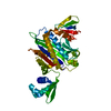
| ||||||||
|---|---|---|---|---|---|---|---|---|---|
| 1 |
| ||||||||
| Unit cell |
|
-Components
| #1: Protein | Mass: 37186.910 Da / Num. of mol.: 1 Source method: isolated from a genetically manipulated source Source: (gene. exp.) Staphylococcus aureus (bacteria) / Strain: ECT-R 2 / Gene: ECTR2_1310 / Production host: References: UniProt: E5R5T0, biotin-[biotin carboxyl-carrier protein] ligase |
|---|---|
| #2: Water | ChemComp-HOH /  Mass: 18.015 Da / Num. of mol.: 212 / Source method: isolated from a natural source / Formula: H2O Mass: 18.015 Da / Num. of mol.: 212 / Source method: isolated from a natural source / Formula: H2O |
-Experimental details
-Experiment
| Experiment | Method: X-RAY DIFFRACTION / Number of used crystals: 1 |
|---|
- Sample preparation
| Crystal | Density Matthews: 2.23 Å3/Da / Density % sol: 44.77 % |
|---|---|
| Crystal grow | Temperature: 293 K / Method: vapor diffusion, hanging drop / pH: 7 Details: Peg 3350, pH 7.0, VAPOR DIFFUSION, HANGING DROP, temperature 293K |
-Data collection
| Diffraction | Mean temperature: 100 K |
|---|---|
| Diffraction source | Source: SYNCHROTRON / Site: Australian Synchrotron  / Beamline: MX1 / Wavelength: 0.95364 Å / Beamline: MX1 / Wavelength: 0.95364 Å |
| Detector | Type: MAR CCD 165 mm / Detector: CCD / Date: Jan 1, 2009 |
| Radiation | Protocol: SINGLE WAVELENGTH / Monochromatic (M) / Laue (L): M / Scattering type: x-ray |
| Radiation wavelength | Wavelength: 0.95364 Å / Relative weight: 1 |
| Reflection | Resolution: 2.1→35 Å / Num. obs: 18537 / % possible obs: 96.1 % / Observed criterion σ(F): 0 / Observed criterion σ(I): 0 |
- Processing
| Software |
| ||||||||||||||||||||||||||||||||||||||||||||||||||||||||||||||||||||||||||||||||||||||||||||||||||||
|---|---|---|---|---|---|---|---|---|---|---|---|---|---|---|---|---|---|---|---|---|---|---|---|---|---|---|---|---|---|---|---|---|---|---|---|---|---|---|---|---|---|---|---|---|---|---|---|---|---|---|---|---|---|---|---|---|---|---|---|---|---|---|---|---|---|---|---|---|---|---|---|---|---|---|---|---|---|---|---|---|---|---|---|---|---|---|---|---|---|---|---|---|---|---|---|---|---|---|---|---|---|
| Refinement | Method to determine structure: MOLECULAR REPLACEMENT Starting model: 3RIR Resolution: 2.1→34.956 Å / SU ML: 0.37 / σ(F): 1.38 / Phase error: 26.65 / Stereochemistry target values: ML
| ||||||||||||||||||||||||||||||||||||||||||||||||||||||||||||||||||||||||||||||||||||||||||||||||||||
| Solvent computation | Shrinkage radii: 0.83 Å / VDW probe radii: 1.1 Å / Solvent model: FLAT BULK SOLVENT MODEL / Bsol: 48.802 Å2 / ksol: 0.386 e/Å3 | ||||||||||||||||||||||||||||||||||||||||||||||||||||||||||||||||||||||||||||||||||||||||||||||||||||
| Displacement parameters |
| ||||||||||||||||||||||||||||||||||||||||||||||||||||||||||||||||||||||||||||||||||||||||||||||||||||
| Refinement step | Cycle: LAST / Resolution: 2.1→34.956 Å
| ||||||||||||||||||||||||||||||||||||||||||||||||||||||||||||||||||||||||||||||||||||||||||||||||||||
| Refine LS restraints |
| ||||||||||||||||||||||||||||||||||||||||||||||||||||||||||||||||||||||||||||||||||||||||||||||||||||
| LS refinement shell | Refine-ID: X-RAY DIFFRACTION / Total num. of bins used: 7
| ||||||||||||||||||||||||||||||||||||||||||||||||||||||||||||||||||||||||||||||||||||||||||||||||||||
| Refinement TLS params. | Method: refined / Refine-ID: X-RAY DIFFRACTION
| ||||||||||||||||||||||||||||||||||||||||||||||||||||||||||||||||||||||||||||||||||||||||||||||||||||
| Refinement TLS group |
|
