 Movie
Movie Controller

[English] 日本語
 Yorodumi
- PDB-1ijx: CRYSTAL STRUCTURE OF THE CYSTEINE-RICH DOMAIN OF SECRETED FRIZZLE... -
+ Open data

- Basic information
| Entry | Database: PDB / ID: 1ijx | ||||||
|---|---|---|---|---|---|---|---|
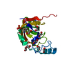
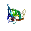
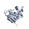
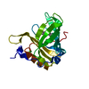
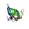
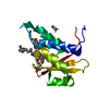
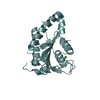
| Title | CRYSTAL STRUCTURE OF THE CYSTEINE-RICH DOMAIN OF SECRETED FRIZZLED-RELATED PROTEIN 3 (SFRP-3;FZB) | ||||||
 Components Components | SECRETED FRIZZLED-RELATED SEQUENCE PROTEIN 3 | ||||||
 Keywords Keywords | SIGNALING PROTEIN / WNT RECEPTOR / FRIZZLED PROTEIN STRUCTURE / CYSTEINE-RICH | ||||||
| Function / homology |  Function and homology information Function and homology informationnegative regulation of hepatocyte differentiation / negative regulation of cell development / convergent extension involved in organogenesis / system development / negative regulation of cartilage development / cochlea morphogenesis / somite development / non-canonical Wnt signaling pathway / Wnt-protein binding / inner ear morphogenesis ...negative regulation of hepatocyte differentiation / negative regulation of cell development / convergent extension involved in organogenesis / system development / negative regulation of cartilage development / cochlea morphogenesis / somite development / non-canonical Wnt signaling pathway / Wnt-protein binding / inner ear morphogenesis / neural crest cell differentiation / negative regulation of Wnt signaling pathway / canonical Wnt signaling pathway / positive regulation of fat cell differentiation / negative regulation of canonical Wnt signaling pathway / negative regulation of cell growth / positive regulation of apoptotic process / negative regulation of cell population proliferation / : / extracellular region / cytoplasm Similarity search - Function | ||||||
| Biological species |  | ||||||
| Method |  X-RAY DIFFRACTION / SYNCHROTRON / MAD / Resolution: 1.9 Å X-RAY DIFFRACTION / SYNCHROTRON / MAD / Resolution: 1.9 Å | ||||||
 Authors Authors | Dann III, C.E. / Hsieh, J.C. / Rattner, A. / Sharma, D. / Nathans, J. / Leahy, D.J. | ||||||
 Citation Citation | Journal: Nature / Year: 2001 Title: Insights into Wnt binding and signalling from the structures of two Frizzled cysteine-rich domains. Authors: Dann III, C.E. / Hsieh, J.C. / Rattner, A. / Sharma, D. / Nathans, J. / Leahy, D.J. | ||||||
| History |
|
- Structure visualization
| Structure viewer | Molecule: MolmilJmol/JSmol |
|---|
- Downloads & links
-Download
| PDBx/mmCIF format | 1ijx.cif.gz | 162 KB | Display | PDBx/mmCIF format |
|---|---|---|---|---|
| PDB format | pdb1ijx.ent.gz | 130.1 KB | Display | PDB format |
| PDBx/mmJSON format | 1ijx.json.gz | Tree view | PDBx/mmJSON format | |
| Others |  Other downloads Other downloads |
-Validation report
| Arichive directory | https://data.pdbj.org/pub/pdb/validation_reports/ij/1ijxftp://data.pdbj.org/pub/pdb/validation_reports/ij/1ijx | HTTPS FTP |
|---|
-Related structure data


-Links
 PDBj
PDBj- Assembly
| Deposited unit | 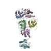
| ||||||||
|---|---|---|---|---|---|---|---|---|---|
| 1 | 
| ||||||||
| 2 | 
| ||||||||
| 3 | 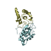
| ||||||||
| Unit cell |
|
-Components
| #1: Protein | Mass: 14199.509 Da / Num. of mol.: 6 / Fragment: CYSTEINE-RICH DOMAIN / Mutation: N17E Source method: isolated from a genetically manipulated source Source: (gene. exp.)  Cricetulus griseus (Chinese hamster) / References: UniProt: P97401 Cricetulus griseus (Chinese hamster) / References: UniProt: P97401#2: Chemical | ChemComp-SO4 / |   Mass: 96.063 Da / Num. of mol.: 1 / Source method: obtained synthetically / Formula: SO4 Mass: 96.063 Da / Num. of mol.: 1 / Source method: obtained synthetically / Formula: SO4#3: Water | ChemComp-HOH / |  Mass: 18.015 Da / Num. of mol.: 499 / Source method: isolated from a natural source / Formula: H2O Mass: 18.015 Da / Num. of mol.: 499 / Source method: isolated from a natural source / Formula: H2OHas protein modification | Y | |
|---|
-Experimental details
-Experiment
| Experiment | Method: X-RAY DIFFRACTION / Number of used crystals: 1 |
|---|
- Sample preparation
| Crystal | Density Matthews: 2.28 Å3/Da / Density % sol: 46.16 % | ||||||||||||||||||||
|---|---|---|---|---|---|---|---|---|---|---|---|---|---|---|---|---|---|---|---|---|---|
| Crystal grow | Temperature: 293 K / Method: vapor diffusion, hanging drop / pH: 6.6 Details: PEG 3350, PEG 400, Sodium Hepes, pH 6.6, VAPOR DIFFUSION, HANGING DROP, temperature 293K | ||||||||||||||||||||
| Crystal grow | *PLUS Temperature: 20 ℃ | ||||||||||||||||||||
| Components of the solutions | *PLUS
|
-Data collection
| Diffraction | Mean temperature: 100 K |
|---|---|
| Diffraction source | Source: SYNCHROTRON / Site: NSLS  / Beamline: X4A / Wavelength: 0.9203 Å / Beamline: X4A / Wavelength: 0.9203 Å |
| Detector | Type: ADSC QUANTUM 4 / Detector: CCD / Date: Jun 11, 2000 |
| Radiation | Monochromator: SAGITALLY FOCUSED Si(111) / Protocol: SINGLE WAVELENGTH / Monochromatic (M) / Laue (L): M / Scattering type: x-ray |
| Radiation wavelength | Wavelength: 0.9203 Å / Relative weight: 1 |
| Reflection | Resolution: 1.9→30 Å / Num. all: 61210 / Num. obs: 61070 / % possible obs: 99.8 % / Observed criterion σ(F): 0 / Observed criterion σ(I): 0 / Redundancy: 7.4 % / Rmerge(I) obs: 0.077 / Net I/σ(I): 26.3 |
| Reflection shell | Resolution: 1.9→1.97 Å / Redundancy: 6.6 % / Rmerge(I) obs: 0.679 / Mean I/σ(I) obs: 2.2 / Num. unique all: 6167 / % possible all: 98.7 |
- Processing
| Software |
| ||||||||||||||||||||||||||||||
|---|---|---|---|---|---|---|---|---|---|---|---|---|---|---|---|---|---|---|---|---|---|---|---|---|---|---|---|---|---|---|---|
| Refinement | Method to determine structure: MAD / Resolution: 1.9→30 Å / σ(F): 0 / σ(I): 0 / Stereochemistry target values: Engh & Huber
| ||||||||||||||||||||||||||||||
| Refinement step | Cycle: LAST / Resolution: 1.9→30 Å
| ||||||||||||||||||||||||||||||
| Software | *PLUS Name: CNS / Version: 1 / Classification: refinement | ||||||||||||||||||||||||||||||
| Refinement | *PLUS Highest resolution: 1.9 Å / Lowest resolution: 30 Å / σ(F): 0 / Rfactor obs: 0.22 / Rfactor Rwork: 0.22 | ||||||||||||||||||||||||||||||
| Solvent computation | *PLUS | ||||||||||||||||||||||||||||||
| Displacement parameters | *PLUS | ||||||||||||||||||||||||||||||
| Refine LS restraints | *PLUS
|
