 Movie
Movie Controller


+ Open data

- Basic information
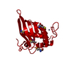
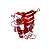
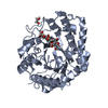
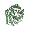
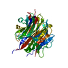
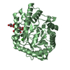
| Entry | Database: PDB / ID: 1j98 | ||||||
|---|---|---|---|---|---|---|---|
| Title | The 1.2 Angstrom Structure of Bacillus subtilis LuxS | ||||||
 Components Components | AUTOINDUCER-2 PRODUCTION PROTEIN LUXS | ||||||
 Keywords Keywords | SIGNALING PROTEIN / Autoinducer synthesis | ||||||
| Function / homology |  Function and homology information Function and homology informationS-ribosylhomocysteine lyase / S-ribosylhomocysteine lyase activity / : / quorum sensing / iron ion binding / cytosol Similarity search - Function | ||||||
| Biological species |  | ||||||
| Method |  X-RAY DIFFRACTION / SYNCHROTRON / MIR / Resolution: 1.2 Å X-RAY DIFFRACTION / SYNCHROTRON / MIR / Resolution: 1.2 Å | ||||||
 Authors Authors | Ruzheinikov, S.N. / Das, S.K. / Sedelnikova, S.E. / Hartley, A. / Foster, S.J. / Horsburgh, M.J. / Cox, A.G. / McCleod, C.W. / Mekhalfia, A. / Blackburn, G.M. ...Ruzheinikov, S.N. / Das, S.K. / Sedelnikova, S.E. / Hartley, A. / Foster, S.J. / Horsburgh, M.J. / Cox, A.G. / McCleod, C.W. / Mekhalfia, A. / Blackburn, G.M. / Rice, D.W. / Baker, P.J. | ||||||
 Citation Citation | Journal: J.Mol.Biol. / Year: 2001 Title: The 1.2 A Structure of a Novel Quorum-Sensing Protein, Bacillus subtilis LuxS Authors: Ruzheinikov, S.N. / Das, S.K. / Sedelnikova, S.E. / Hartley, A. / Foster, S.J. / Horsburgh, M.J. / Cox, A.G. / McCleod, C.W. / Mekhalfia, A. / Blackburn, G.M. / Rice, D.W. / Baker, P.J. #1: Journal: Acta Crystallogr.,Sect.D / Year: 2001Title: Cloning, Purification, Crystallization and Preliminary Crystallographic Analysis of Bacillus subtilis LuxS Authors: Das, S.K. / Sedelnikova, S.E. / Baker, P.J. / Ruzheinikov, S.N. / Foster, S.J. / Hartley, A. / Horsburgh, M.J. / Rice, D.W. | ||||||
| History |
|
- Structure visualization
| Structure viewer | Molecule: MolmilJmol/JSmol |
|---|
- Downloads & links
-Download
| PDBx/mmCIF format | 1j98.cif.gz | 81.2 KB | Display | PDBx/mmCIF format |
|---|---|---|---|---|
| PDB format | pdb1j98.ent.gz | 65.1 KB | Display | PDB format |
| PDBx/mmJSON format | 1j98.json.gz | Tree view | PDBx/mmJSON format | |
| Others |  Other downloads Other downloads |
-Validation report
| Arichive directory | https://data.pdbj.org/pub/pdb/validation_reports/j9/1j98ftp://data.pdbj.org/pub/pdb/validation_reports/j9/1j98 | HTTPS FTP |
|---|
-Related structure data


-Links
 PDBj
PDBj

- Assembly
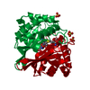
| Deposited unit | 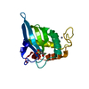
| ||||||||
|---|---|---|---|---|---|---|---|---|---|
| 1 | 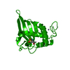
| ||||||||
| Unit cell |
| ||||||||
| Details | The second part of the biological assembly is generated by the two fold axis: x, x-y, 5/6-z. |
-Components
| #1: Protein | Mass: 17790.219 Da / Num. of mol.: 1 / Mutation: P96T Source method: isolated from a genetically manipulated source Source: (gene. exp.) |
|---|---|
| #2: Chemical | ChemComp-ZN /   Mass: 65.409 Da / Num. of mol.: 1 / Source method: obtained synthetically / Formula: Zn Mass: 65.409 Da / Num. of mol.: 1 / Source method: obtained synthetically / Formula: Zn |
| #3: Water | ChemComp-HOH /  Mass: 18.015 Da / Num. of mol.: 217 / Source method: isolated from a natural source / Formula: H2O Mass: 18.015 Da / Num. of mol.: 217 / Source method: isolated from a natural source / Formula: H2O |
-Experimental details
-Experiment
| Experiment | Method: X-RAY DIFFRACTION / Number of used crystals: 1 |
|---|
- Sample preparation
| Crystal | Density Matthews: 2.5 Å3/Da / Density % sol: 39.56 % | |||||||||||||||
|---|---|---|---|---|---|---|---|---|---|---|---|---|---|---|---|---|
| Crystal grow | Temperature: 290 K / Method: vapor diffusion, hanging drop / pH: 8 Details: 1.8 - 2.4M Ammonium sulphate, 0.1M Tris-HCl, pH 8.0, VAPOR DIFFUSION, HANGING DROP, temperature 290K | |||||||||||||||
| Crystal grow | *PLUS | |||||||||||||||
| Components of the solutions | *PLUS
|
-Data collection
| Diffraction | Mean temperature: 100 K |
|---|---|
| Diffraction source | Source: SYNCHROTRON / Site: SRS  / Beamline: PX14.2 / Wavelength: 0.978 Å / Beamline: PX14.2 / Wavelength: 0.978 Å |
| Detector | Type: ADSC QUANTUM 4 / Detector: CCD / Date: Apr 9, 2001 |
| Radiation | Protocol: SINGLE WAVELENGTH / Monochromatic (M) / Laue (L): M / Scattering type: x-ray |
| Radiation wavelength | Wavelength: 0.978 Å / Relative weight: 1 |
| Reflection | Resolution: 1.2→10 Å / Num. all: 53328 / Num. obs: 53328 / % possible obs: 96.5 % / Observed criterion σ(F): 0 / Observed criterion σ(I): 0 / Redundancy: 5.5 % / Biso Wilson estimate: 13.6 Å2 / Rmerge(I) obs: 0.048 / Net I/σ(I): 32.9 |
| Reflection shell | Resolution: 1.2→1.22 Å / Redundancy: 2.7 % / Rmerge(I) obs: 0.257 / Mean I/σ(I) obs: 2.8 / Num. unique all: 1714 / % possible all: 63.3 |
| Reflection | *PLUS Num. measured all: 288300 |
| Reflection shell | *PLUS % possible obs: 63.6 % |
- Processing
| Software |
| |||||||||||||||||||||||||
|---|---|---|---|---|---|---|---|---|---|---|---|---|---|---|---|---|---|---|---|---|---|---|---|---|---|---|
| Refinement | Method to determine structure: MIR / Resolution: 1.2→10 Å / SU B: 1.056 / SU ML: 0.025 / Isotropic thermal model: anisotropic / Cross valid method: THROUGHOUT / σ(F): 0 / σ(I): 0 / ESU R: 0.031 / ESU R Free: 0.031 / Details: HYDROGENS HAVE BEEN ADDED IN THE RIDING POSITIONS
| |||||||||||||||||||||||||
| Displacement parameters | Biso mean: 16.8 Å2
| |||||||||||||||||||||||||
| Refine analyze |
| |||||||||||||||||||||||||
| Refinement step | Cycle: LAST / Resolution: 1.2→10 Å
| |||||||||||||||||||||||||
| Refine LS restraints |
| |||||||||||||||||||||||||
| Software | *PLUS Name: REFMAC / Classification: refinement | |||||||||||||||||||||||||
| Refinement | *PLUS Highest resolution: 1.2 Å / σ(F): 0 / % reflection Rfree: 5.1 % / Rfactor obs: 0.126 | |||||||||||||||||||||||||
| Solvent computation | *PLUS | |||||||||||||||||||||||||
| Displacement parameters | *PLUS Biso mean: 16.8 Å2 |
