 Movie
Movie Controller

[English] 日本語
 Yorodumi
- PDB-1jqw: THE 2.3 ANGSTROM RESOLUTION STRUCTURE OF BACILLUS SUBTILIS LUXS/H... -
+ Open data

- Basic information
| Entry | Database: PDB / ID: 1jqw | ||||||
|---|---|---|---|---|---|---|---|
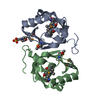
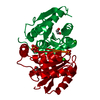
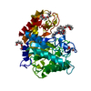
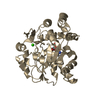
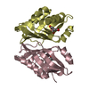
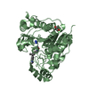
| Title | THE 2.3 ANGSTROM RESOLUTION STRUCTURE OF BACILLUS SUBTILIS LUXS/HOMOCYSTEINE COMPLEX | ||||||
 Components Components | Autoinducer-2 production protein luxS | ||||||
 Keywords Keywords | SIGNALING PROTEIN / AUTOINDUCER SYNTHESIS | ||||||
| Function / homology |  Function and homology information Function and homology informationS-ribosylhomocysteine lyase / S-ribosylhomocysteine lyase activity / : / quorum sensing / iron ion binding / cytosol Similarity search - Function | ||||||
| Biological species |  | ||||||
| Method |  X-RAY DIFFRACTION / MOLECULAR REPLACEMENT / Resolution: 2.3 Å X-RAY DIFFRACTION / MOLECULAR REPLACEMENT / Resolution: 2.3 Å | ||||||
 Authors Authors | Ruzheinikov, S.N. / Das, S.K. / Sedelnikova, S.E. / Hartley, A. / Foster, S.J. / Horsburgh, M.J. / Cox, A.G. / McCleod, C.W. / Mekhalfia, A. / Blackburn, G.M. ...Ruzheinikov, S.N. / Das, S.K. / Sedelnikova, S.E. / Hartley, A. / Foster, S.J. / Horsburgh, M.J. / Cox, A.G. / McCleod, C.W. / Mekhalfia, A. / Blackburn, G.M. / Rice, D.W. / Baker, P.J. | ||||||
 Citation Citation | Journal: J.Mol.Biol. / Year: 2001 Title: The 1.2 A structure of a novel quorum-sensing protein, Bacillus subtilis LuxS Authors: Ruzheinikov, S.N. / Das, S.K. / Sedelnikova, S.E. / Hartley, A. / Foster, S.J. / Horsburgh, M.J. / Cox, A.G. / McCleod, C.W. / Mekhalfia, A. / Blackburn, G.M. / Rice, D.W. / Baker, P.J. #1: Journal: TO BE PUBLISHEDTitle: PURIFICATION, CRYSTALLIZATION AND PRELIMINARY CRYSTALLOGRAPHIC ANALYSIS OF BACILLUS SUBTILIS LUXS Authors: Das, S.K. / Sedelnikova, S.E. / Baker, P.J. / Ruzheinikov, S.N. / Foster, S.J. / Hartley, A. / Horsburgh, M.J. / Rice, D.W. | ||||||
| History |
|
- Structure visualization
| Structure viewer | Molecule: MolmilJmol/JSmol |
|---|
- Downloads & links
-Download
| PDBx/mmCIF format | 1jqw.cif.gz | 45.9 KB | Display | PDBx/mmCIF format |
|---|---|---|---|---|
| PDB format | pdb1jqw.ent.gz | 32.2 KB | Display | PDB format |
| PDBx/mmJSON format | 1jqw.json.gz | Tree view | PDBx/mmJSON format | |
| Others |  Other downloads Other downloads |
-Validation report
| Arichive directory | https://data.pdbj.org/pub/pdb/validation_reports/jq/1jqwftp://data.pdbj.org/pub/pdb/validation_reports/jq/1jqw | HTTPS FTP |
|---|
-Related structure data
| Related structure data |  1j98SC  1jviC S: Starting model for refinement C: citing same article ( |
|---|---|
| Similar structure data |

-Links
 PDBj
PDBj

- Assembly
| Deposited unit | 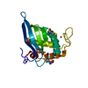
| ||||||||
|---|---|---|---|---|---|---|---|---|---|
| 1 | 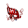
| ||||||||
| Unit cell |
| ||||||||
| Details | The biological assembly is a dimer generated from the monomer in the asymmetric unit by the matrix: BIOMT1 2 0.500000 0.866025 0.000000 -31.39000 BIOMT2 2 0.866025 -0.500000 0.000000 54.36907 BIOMT3 2 0.000000 0.000000 -1.000000 -24.93000 |
-Components
| #1: Protein | Mass: 17790.219 Da / Num. of mol.: 1 / Mutation: P96T Source method: isolated from a genetically manipulated source Source: (gene. exp.) | ||
|---|---|---|---|
| #2: Chemical | ChemComp-ZN /   Mass: 65.409 Da / Num. of mol.: 1 / Source method: obtained synthetically / Formula: Zn Mass: 65.409 Da / Num. of mol.: 1 / Source method: obtained synthetically / Formula: Zn | ||
| #3: Chemical |   Type: L-peptide linking / Mass: 135.185 Da / Num. of mol.: 3 / Source method: obtained synthetically / Formula: C4H9NO2S Type: L-peptide linking / Mass: 135.185 Da / Num. of mol.: 3 / Source method: obtained synthetically / Formula: C4H9NO2S#4: Water | ChemComp-HOH / |  Mass: 18.015 Da / Num. of mol.: 53 / Source method: isolated from a natural source / Formula: H2O Mass: 18.015 Da / Num. of mol.: 53 / Source method: isolated from a natural source / Formula: H2O |
-Experimental details
-Experiment
| Experiment | Method: X-RAY DIFFRACTION / Number of used crystals: 1 |
|---|
- Sample preparation
| Crystal | Density Matthews: 2.28 Å3/Da / Density % sol: 46.05 % | |||||||||||||||
|---|---|---|---|---|---|---|---|---|---|---|---|---|---|---|---|---|
| Crystal grow | Temperature: 290 K / Method: vapor diffusion, hanging drop / pH: 8 Details: 1.8 - 2.4M AMMONIUM SULPHATE, 0.1M TRIS-HCL, pH 8.0, VAPOR DIFFUSION, HANGING DROP, temperature 290K | |||||||||||||||
| Crystal grow | *PLUS | |||||||||||||||
| Components of the solutions | *PLUS
|
-Data collection
| Diffraction | Mean temperature: 100 K |
|---|---|
| Diffraction source | Source: ROTATING ANODE / Type: RIGAKU RU200 / Wavelength: 1.5418 Å |
| Radiation | Protocol: SINGLE WAVELENGTH / Monochromatic (M) / Laue (L): M / Scattering type: x-ray |
| Radiation wavelength | Wavelength: 1.5418 Å / Relative weight: 1 |
| Reflection | Resolution: 2.3→20 Å / Num. all: 7264 / Num. obs: 7264 / % possible obs: 90.8 % / Observed criterion σ(F): 0 / Observed criterion σ(I): 0 / Redundancy: 5.36 % / Rmerge(I) obs: 0.044 / Net I/σ(I): 20.8 |
| Reflection shell | Resolution: 2.3→2.35 Å / Rmerge(I) obs: 0.378 / Mean I/σ(I) obs: 3.35 / Num. unique all: 494 / % possible all: 94.5 |
| Reflection | *PLUS Num. measured all: 38530 / Rmerge(I) obs: 0.062 |
| Reflection shell | *PLUS % possible obs: 94.5 % / Num. unique obs: 185 / Rmerge(I) obs: 0.501 |
- Processing
| Software |
| |||||||||||||||||||||||||||
|---|---|---|---|---|---|---|---|---|---|---|---|---|---|---|---|---|---|---|---|---|---|---|---|---|---|---|---|---|
| Refinement | Method to determine structure: MOLECULAR REPLACEMENT Starting model: 1J98 Resolution: 2.3→10 Å / Isotropic thermal model: Isotropic / Cross valid method: THROUGHOUT / σ(F): 1 / Stereochemistry target values: Engh & Huber
| |||||||||||||||||||||||||||
| Displacement parameters | Biso mean: 44.52 Å2 | |||||||||||||||||||||||||||
| Refine analyze |
| |||||||||||||||||||||||||||
| Refinement step | Cycle: LAST / Resolution: 2.3→10 Å
| |||||||||||||||||||||||||||
| Refine LS restraints |
| |||||||||||||||||||||||||||
| Software | *PLUS Name: CNS / Version: 1 / Classification: refinement | |||||||||||||||||||||||||||
| Refinement | *PLUS Highest resolution: 2.3 Å / Lowest resolution: 10 Å / σ(F): 1 / Rfactor obs: 0.175 | |||||||||||||||||||||||||||
| Solvent computation | *PLUS | |||||||||||||||||||||||||||
| Displacement parameters | *PLUS | |||||||||||||||||||||||||||
| Refine LS restraints | *PLUS
|
