 Movie
Movie Controller

[English] 日本語
 Yorodumi
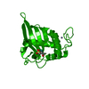
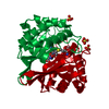
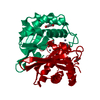
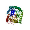
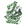
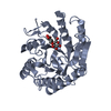
- PDB-1jvi: THE 2.2 ANGSTROM RESOLUTION STRUCTURE OF BACILLUS SUBTILIS LUXS/R... -
+ Open data

- Basic information
| Entry | Database: PDB / ID: 1jvi | |||||||||
|---|---|---|---|---|---|---|---|---|---|---|
| Title | THE 2.2 ANGSTROM RESOLUTION STRUCTURE OF BACILLUS SUBTILIS LUXS/RIBOSILHOMOCYSTEINE COMPLEX | |||||||||
 Components Components | Autoinducer-2 production protein luxS | |||||||||
 Keywords Keywords | SIGNALING PROTEIN / AUTOINDUCER SYNTHESIS | |||||||||
| Function / homology |  Function and homology information Function and homology informationS-ribosylhomocysteine lyase / S-ribosylhomocysteine lyase activity / : / quorum sensing / iron ion binding / cytosol Similarity search - Function | |||||||||
| Biological species |  | |||||||||
| Method |  X-RAY DIFFRACTION / MOLECULAR REPLACEMENT / Resolution: 2.2 Å X-RAY DIFFRACTION / MOLECULAR REPLACEMENT / Resolution: 2.2 Å | |||||||||
 Authors Authors | Ruzheinikov, S.N. / Das, S.K. / Sedelnikova, S.E. / Hartley, A. / Foster, S.J. / Horsburgh, M.J. / Cox, A.G. / McCleod, C.W. / Mekhalfia, A. / Blackburn, G.M. ...Ruzheinikov, S.N. / Das, S.K. / Sedelnikova, S.E. / Hartley, A. / Foster, S.J. / Horsburgh, M.J. / Cox, A.G. / McCleod, C.W. / Mekhalfia, A. / Blackburn, G.M. / Rice, D.W. / Baker, P.J. | |||||||||
 Citation Citation | Journal: J.Mol.Biol. / Year: 2001 Title: The 1.2 A structure of a novel quorum-sensing protein, Bacillus subtilis LuxS Authors: Ruzheinikov, S.N. / Das, S.K. / Sedelnikova, S.E. / Hartley, A. / Foster, S.J. / Horsburgh, M.J. / Cox, A.G. / McCleod, C.W. / Mekhalfia, A. / Blackburn, G.M. / Rice, D.W. / Baker, P.J. #1: Journal: Acta Crystallogr.,Sect.D / Year: 2001Title: Cloning, purification, crystallization and preliminary crystallographic analysis of Bacillus subtilis LuxS. Authors: Das, S.K. / Sedelnikova, S.E. / Baker, P.J. / Ruzheinikov, S.N. / Foster, S. / Hartley, A. / Horsburg, M.J. / Rice, D.W. | |||||||||
| History |
|
- Structure visualization
| Structure viewer | Molecule: MolmilJmol/JSmol |
|---|
- Downloads & links
-Download
| PDBx/mmCIF format | 1jvi.cif.gz | 47.7 KB | Display | PDBx/mmCIF format |
|---|---|---|---|---|
| PDB format | pdb1jvi.ent.gz | 33.1 KB | Display | PDB format |
| PDBx/mmJSON format | 1jvi.json.gz | Tree view | PDBx/mmJSON format | |
| Others |  Other downloads Other downloads |
-Validation report
| Arichive directory | https://data.pdbj.org/pub/pdb/validation_reports/jv/1jviftp://data.pdbj.org/pub/pdb/validation_reports/jv/1jvi | HTTPS FTP |
|---|
-Related structure data
| Related structure data |  1j98SC 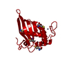 1jqwC S: Starting model for refinement C: citing same article ( |
|---|---|
| Similar structure data |


-Links
 PDBj
PDBj

- Assembly
| Deposited unit | 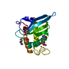
| ||||||||
|---|---|---|---|---|---|---|---|---|---|
| 1 | 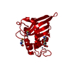
| ||||||||
| Unit cell |
| ||||||||
| Details | The second part of the biological assembly is generated by the two fold axis: X, X-Y, 5/6-Z |
-Components
-Protein / Sugars , 2 types, 2 molecules A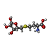
| #1: Protein | Mass: 17790.219 Da / Num. of mol.: 1 / Mutation: P96T Source method: isolated from a genetically manipulated source Source: (gene. exp.) |
|---|---|
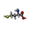
| #4: Sugar | ChemComp-RHC / ( Type: D-saccharide, beta linking / Mass: 267.299 Da / Num. of mol.: 1 / Source method: obtained synthetically / Formula: C9H17NO6S Type: D-saccharide, beta linking / Mass: 267.299 Da / Num. of mol.: 1 / Source method: obtained synthetically / Formula: C9H17NO6S |
-Non-polymers , 4 types, 84 molecules 


| #2: Chemical | ChemComp-ZN /  Mass: 65.409 Da / Num. of mol.: 1 / Source method: obtained synthetically / Formula: Zn Mass: 65.409 Da / Num. of mol.: 1 / Source method: obtained synthetically / Formula: Zn |
|---|---|
| #3: Chemical | ChemComp-SO4 /  Mass: 96.063 Da / Num. of mol.: 1 / Source method: obtained synthetically / Formula: SO4 Mass: 96.063 Da / Num. of mol.: 1 / Source method: obtained synthetically / Formula: SO4 |
| #5: Chemical | ChemComp-HCS /  Type: L-peptide linking / Mass: 135.185 Da / Num. of mol.: 1 / Source method: obtained synthetically / Formula: C4H9NO2S Type: L-peptide linking / Mass: 135.185 Da / Num. of mol.: 1 / Source method: obtained synthetically / Formula: C4H9NO2S |
| #6: Water | ChemComp-HOH / Mass: 18.015 Da / Num. of mol.: 81 / Source method: isolated from a natural source / Formula: H2O |
-Experimental details
-Experiment
| Experiment | Method: X-RAY DIFFRACTION / Number of used crystals: 1 |
|---|
- Sample preparation
| Crystal | Density Matthews: 2.55 Å3/Da / Density % sol: 45.3 % | |||||||||||||||
|---|---|---|---|---|---|---|---|---|---|---|---|---|---|---|---|---|
| Crystal grow | Temperature: 290 K / Method: vapor diffusion, hanging drop Details: 1.8 - 2.4M AMMONIUM SULPHATE, 0.1M TRIS-HCL, VAPOR DIFFUSION, HANGING DROP, temperature 290K | |||||||||||||||
| Crystal grow | *PLUS pH: 8 | |||||||||||||||
| Components of the solutions | *PLUS
|
-Data collection
| Diffraction | Mean temperature: 100 K |
|---|---|
| Diffraction source | Source: ROTATING ANODE / Type: RIGAKU / Wavelength: 1.5418 Å |
| Detector | Type: MARRESEARCH / Detector: IMAGE PLATE |
| Radiation | Protocol: SINGLE WAVELENGTH / Monochromatic (M) / Laue (L): M / Scattering type: x-ray |
| Radiation wavelength | Wavelength: 1.5418 Å / Relative weight: 1 |
| Reflection | Resolution: 2.2→20 Å / Num. all: 8944 / Num. obs: 8944 / % possible obs: 95.7 % / Observed criterion σ(F): 0 / Observed criterion σ(I): 0 / Redundancy: 2.63 % / Biso Wilson estimate: 36.49 Å2 / Rmerge(I) obs: 0.062 / Net I/σ(I): 13.1 |
| Reflection shell | Resolution: 2.2→2.25 Å / Rmerge(I) obs: 0.36 / Num. unique all: 553 / % possible all: 92.5 |
| Reflection | *PLUS Num. measured all: 24877 |
| Reflection shell | *PLUS Num. unique obs: 339 / Rmerge(I) obs: 0.36 / Mean I/σ(I) obs: 2.3 |
- Processing
| Software |
| ||||||||||||||||||||||||||||||||||||||||||
|---|---|---|---|---|---|---|---|---|---|---|---|---|---|---|---|---|---|---|---|---|---|---|---|---|---|---|---|---|---|---|---|---|---|---|---|---|---|---|---|---|---|---|---|
| Refinement | Method to determine structure: MOLECULAR REPLACEMENT Starting model: PDB ENTRY 1J98 Resolution: 2.2→10 Å / Isotropic thermal model: Isotropic / Cross valid method: THROUGHOUT / σ(F): 1 / Stereochemistry target values: Engh & Huber
| ||||||||||||||||||||||||||||||||||||||||||
| Displacement parameters | Biso mean: 37.37 Å2 | ||||||||||||||||||||||||||||||||||||||||||
| Refine analyze |
| ||||||||||||||||||||||||||||||||||||||||||
| Refinement step | Cycle: LAST / Resolution: 2.2→10 Å
| ||||||||||||||||||||||||||||||||||||||||||
| Refine LS restraints |
| ||||||||||||||||||||||||||||||||||||||||||
| LS refinement shell |
| ||||||||||||||||||||||||||||||||||||||||||
| Software | *PLUS Name: CNS / Version: 1 / Classification: refinement | ||||||||||||||||||||||||||||||||||||||||||
| Refinement | *PLUS Highest resolution: 2.2 Å / Lowest resolution: 10 Å / σ(F): 1 / Rfactor obs: 0.174 | ||||||||||||||||||||||||||||||||||||||||||
| Solvent computation | *PLUS | ||||||||||||||||||||||||||||||||||||||||||
| Displacement parameters | *PLUS | ||||||||||||||||||||||||||||||||||||||||||
| Refine LS restraints | *PLUS
| ||||||||||||||||||||||||||||||||||||||||||
| LS refinement shell | *PLUS Lowest resolution: 10 Å / Rfactor Rfree: 0.212 / Rfactor Rwork: 0.142 |
