 Movie
Movie Controller

[English] 日本語
 Yorodumi
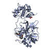
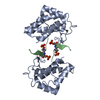
- PDB-1ijy: CRYSTAL STRUCTURE OF THE CYSTEINE-RICH DOMAIN OF MOUSE FRIZZLED 8... -
+ Open data

- Basic information
| Entry | Database: PDB / ID: 1ijy | ||||||
|---|---|---|---|---|---|---|---|
| Title | CRYSTAL STRUCTURE OF THE CYSTEINE-RICH DOMAIN OF MOUSE FRIZZLED 8 (MFZ8) | ||||||
 Components Components | FRIZZLED HOMOLOG 8 | ||||||
 Keywords Keywords | SIGNALING PROTEIN / WNT RECEPTOR / FRIZZLED PROTEIN STRUCTURE / CYSTEINE-RICH | ||||||
| Function / homology |  Function and homology information Function and homology informationWnt-Frizzled-LRP5/6 complex / Asymmetric localization of PCP proteins / Regulation of FZD by ubiquitination / Wnt receptor activity / non-canonical Wnt signaling pathway / Wnt-protein binding / canonical Wnt signaling pathway / neuronal dense core vesicle / PDZ domain binding / G protein-coupled receptor activity ...Wnt-Frizzled-LRP5/6 complex / Asymmetric localization of PCP proteins / Regulation of FZD by ubiquitination / Wnt receptor activity / non-canonical Wnt signaling pathway / Wnt-protein binding / canonical Wnt signaling pathway / neuronal dense core vesicle / PDZ domain binding / G protein-coupled receptor activity / Wnt signaling pathway / T cell differentiation in thymus / angiogenesis / signaling receptor binding / ubiquitin protein ligase binding / Golgi apparatus / extracellular region / plasma membrane Similarity search - Function | ||||||
| Biological species |  | ||||||
| Method |  X-RAY DIFFRACTION / SYNCHROTRON / MOLECULAR REPLACEMENT / Resolution: 1.35 Å X-RAY DIFFRACTION / SYNCHROTRON / MOLECULAR REPLACEMENT / Resolution: 1.35 Å | ||||||
 Authors Authors | Dann III, C.E. / Hsieh, J.C. / Rattner, A. / Sharma, D. / Nathans, J. / Leahy, D.J. | ||||||
 Citation Citation | Journal: Nature / Year: 2001 Title: Insights into Wnt binding and signalling from the structures of two Frizzled cysteine-rich domains. Authors: Dann III, C.E. / Hsieh, J.C. / Rattner, A. / Sharma, D. / Nathans, J. / Leahy, D.J. | ||||||
| History |
|
- Structure visualization
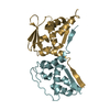
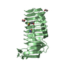
| Structure viewer | Molecule: MolmilJmol/JSmol |
|---|
- Downloads & links
-Download
| PDBx/mmCIF format | 1ijy.cif.gz | 66.9 KB | Display | PDBx/mmCIF format |
|---|---|---|---|---|
| PDB format | pdb1ijy.ent.gz | 49.3 KB | Display | PDB format |
| PDBx/mmJSON format | 1ijy.json.gz | Tree view | PDBx/mmJSON format | |
| Others |  Other downloads Other downloads |
-Validation report
| Arichive directory | https://data.pdbj.org/pub/pdb/validation_reports/ij/1ijyftp://data.pdbj.org/pub/pdb/validation_reports/ij/1ijy | HTTPS FTP |
|---|
-Related structure data
| Related structure data | 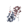 1ijxSC S: Starting model for refinement C: citing same article ( |
|---|---|
| Similar structure data |


-Links
 PDBj
PDBj

- Assembly
| Deposited unit | 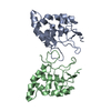
| ||||||||
|---|---|---|---|---|---|---|---|---|---|
| 1 |
| ||||||||
| Unit cell |
|
-Components
| #1: Protein | Mass: 14943.059 Da / Num. of mol.: 2 / Fragment: CYSTEINE-RICH DOMAIN / Mutation: N22E, N125E Source method: isolated from a genetically manipulated source Source: (gene. exp.)  Cricetulus griseus (Chinese hamster) / References: UniProt: Q61091 Cricetulus griseus (Chinese hamster) / References: UniProt: Q61091#2: Water | ChemComp-HOH / |  Mass: 18.015 Da / Num. of mol.: 279 / Source method: isolated from a natural source / Formula: H2O Mass: 18.015 Da / Num. of mol.: 279 / Source method: isolated from a natural source / Formula: H2OHas protein modification | Y | |
|---|
-Experimental details
-Experiment
| Experiment | Method: X-RAY DIFFRACTION / Number of used crystals: 1 |
|---|
- Sample preparation
| Crystal | Density Matthews: 1.94 Å3/Da / Density % sol: 36.55 % | |||||||||||||||
|---|---|---|---|---|---|---|---|---|---|---|---|---|---|---|---|---|
| Crystal grow | Temperature: 293 K / Method: vapor diffusion, hanging drop / pH: 7.3 Details: Ammonium Sulfate, Hepes, pH 7.3, VAPOR DIFFUSION, HANGING DROP, temperature 293K | |||||||||||||||
| Crystal grow | *PLUS | |||||||||||||||
| Components of the solutions | *PLUS
|
-Data collection
| Diffraction | Mean temperature: 100 K |
|---|---|
| Diffraction source | Source: SYNCHROTRON / Site: NSLS  / Beamline: X4A / Wavelength: 0.9197 Å / Beamline: X4A / Wavelength: 0.9197 Å |
| Detector | Type: ADSC QUANTUM 4 / Detector: CCD / Date: Mar 5, 2001 |
| Radiation | Monochromator: SAGITALLY FOCUSED Si(111) / Protocol: SINGLE WAVELENGTH / Monochromatic (M) / Laue (L): M / Scattering type: x-ray |
| Radiation wavelength | Wavelength: 0.9197 Å / Relative weight: 1 |
| Reflection | Resolution: 1.35→30 Å / Num. all: 50707 / Num. obs: 48062 / % possible obs: 94.8 % / Observed criterion σ(F): 0 / Observed criterion σ(I): 0 / Redundancy: 2.8 % / Rmerge(I) obs: 0.051 / Net I/σ(I): 20.5 |
| Reflection shell | Resolution: 1.35→1.4 Å / Redundancy: 2.8 % / Rmerge(I) obs: 0.437 / Mean I/σ(I) obs: 2.1 / Num. unique all: 4993 / % possible all: 99.4 |
- Processing
| Software |
| ||||||||||||||||||||||||||||||
|---|---|---|---|---|---|---|---|---|---|---|---|---|---|---|---|---|---|---|---|---|---|---|---|---|---|---|---|---|---|---|---|
| Refinement | Method to determine structure: MOLECULAR REPLACEMENT Starting model: PDB ENTRY 1IJX Resolution: 1.35→30 Å / σ(F): 0 / σ(I): 0 / Stereochemistry target values: Engh & Huber
| ||||||||||||||||||||||||||||||
| Refinement step | Cycle: LAST / Resolution: 1.35→30 Å
| ||||||||||||||||||||||||||||||
| Software | *PLUS Name: CNS / Classification: refinement | ||||||||||||||||||||||||||||||
| Refinement | *PLUS Lowest resolution: 30 Å / σ(F): 0 / Rfactor obs: 0.225 | ||||||||||||||||||||||||||||||
| Solvent computation | *PLUS | ||||||||||||||||||||||||||||||
| Displacement parameters | *PLUS | ||||||||||||||||||||||||||||||
| Refine LS restraints | *PLUS
|
