 Movie
Movie Controller

[English] 日本語
 Yorodumi
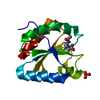
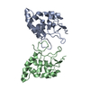
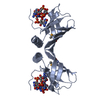
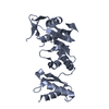
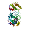
- PDB-5nyo: Crystal structure of an atypical poplar thioredoxin-like2.1 varia... -
+ Open data

- Basic information
| Entry | Database: PDB / ID: 5nyo | |||||||||
|---|---|---|---|---|---|---|---|---|---|---|
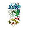
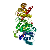
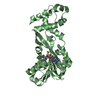
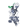
| Title | Crystal structure of an atypical poplar thioredoxin-like2.1 variant in dimeric form | |||||||||
 Components Components | Thioredoxin-like protein 2.1 | |||||||||
 Keywords Keywords | OXIDOREDUCTASE / atypical thioredoxin / disulfide exchange / oxidoredutase | |||||||||
| Function / homology | Thioredoxin-like 3-1/3-2 / chloroplast stroma / Thioredoxin / Thioredoxin domain profile. / Thioredoxin domain / Thioredoxin-like superfamily / Thioredoxin-like protein 2.1 Function and homology information Function and homology information | |||||||||
| Biological species |  | |||||||||
| Method |  X-RAY DIFFRACTION / SYNCHROTRON / MOLECULAR REPLACEMENT / Resolution: 2.25 Å X-RAY DIFFRACTION / SYNCHROTRON / MOLECULAR REPLACEMENT / Resolution: 2.25 Å | |||||||||
 Authors Authors | Chibani, K. / Saul, F.A. / Haouz, A. / Rouhier, N. | |||||||||
| Funding support |  France, 2items France, 2items
| |||||||||
 Citation Citation | Journal: FEBS Lett. / Year: 2018 Title: Structural snapshots along the reaction mechanism of the atypical poplar thioredoxin-like2.1. Authors: Chibani, K. / Saul, F. / Didierjean, C. / Rouhier, N. / Haouz, A. | |||||||||
| History |
|
- Structure visualization
| Structure viewer | Molecule: MolmilJmol/JSmol |
|---|
- Downloads & links
-Download
| PDBx/mmCIF format | 5nyo.cif.gz | 113.8 KB | Display | PDBx/mmCIF format |
|---|---|---|---|---|
| PDB format | pdb5nyo.ent.gz | 87.6 KB | Display | PDB format |
| PDBx/mmJSON format | 5nyo.json.gz | Tree view | PDBx/mmJSON format | |
| Others |  Other downloads Other downloads |
-Validation report
| Arichive directory | https://data.pdbj.org/pub/pdb/validation_reports/ny/5nyoftp://data.pdbj.org/pub/pdb/validation_reports/ny/5nyo | HTTPS FTP |
|---|
-Related structure data


-Links
 PDBj
PDBj

- Assembly
| Deposited unit | 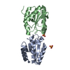
| ||||||||
|---|---|---|---|---|---|---|---|---|---|
| 1 |
| ||||||||
| Unit cell |
|
-Components
| #1: Protein | Mass: 14158.645 Da / Num. of mol.: 2 / Mutation: C45S Engineered mutation Source method: isolated from a genetically manipulated source Source: (gene. exp.) Plasmid: pET3d / Details (production host): T7 expression system / Production host:  #2: Chemical |   Mass: 96.063 Da / Num. of mol.: 2 / Source method: obtained synthetically / Formula: SO4 Mass: 96.063 Da / Num. of mol.: 2 / Source method: obtained synthetically / Formula: SO4#3: Water | ChemComp-HOH / |  Mass: 18.015 Da / Num. of mol.: 98 / Source method: isolated from a natural source / Formula: H2O Mass: 18.015 Da / Num. of mol.: 98 / Source method: isolated from a natural source / Formula: H2OHas protein modification | Y | |
|---|
-Experimental details
-Experiment
| Experiment | Method: X-RAY DIFFRACTION / Number of used crystals: 1 |
|---|
- Sample preparation
| Crystal | Density Matthews: 2.4 Å3/Da / Density % sol: 48.75 % |
|---|---|
| Crystal grow | Temperature: 291 K / Method: vapor diffusion, sitting drop / Details: 0.2M sodium formate, 20% (w/v) PEG3350 |
-Data collection
| Diffraction | Mean temperature: 110 K |
|---|---|
| Diffraction source | Source: SYNCHROTRON / Site: SOLEIL / Beamline: PROXIMA 1 / Wavelength: 0.98011 Å |
| Detector | Type: DECTRIS PILATUS 6M / Detector: PIXEL / Date: Sep 16, 2016 |
| Radiation | Protocol: SINGLE WAVELENGTH / Monochromatic (M) / Laue (L): M / Scattering type: x-ray |
| Radiation wavelength | Wavelength: 0.98011 Å / Relative weight: 1 |
| Reflection | Resolution: 2.25→46.4 Å / Num. obs: 13773 / % possible obs: 100 % / Redundancy: 8.5 % / Biso Wilson estimate: 62.95 Å2 / CC1/2: 0.998 / Rmerge(I) obs: 0.068 / Rpim(I) all: 0.035 / Net I/σ(I): 14.9 |
| Reflection shell | Resolution: 2.25→2.32 Å / Redundancy: 8.8 % / Rmerge(I) obs: 1.078 / Mean I/σ(I) obs: 2.1 / Num. unique obs: 1242 / CC1/2: 0.745 / Rpim(I) all: 0.552 / % possible all: 100 |
- Processing
| Software |
| ||||||||||||||||||||||||||||||||||||||||||||||||||||||||||||||||||||||||||||||||||||||||||||||||||||||||||||||||||
|---|---|---|---|---|---|---|---|---|---|---|---|---|---|---|---|---|---|---|---|---|---|---|---|---|---|---|---|---|---|---|---|---|---|---|---|---|---|---|---|---|---|---|---|---|---|---|---|---|---|---|---|---|---|---|---|---|---|---|---|---|---|---|---|---|---|---|---|---|---|---|---|---|---|---|---|---|---|---|---|---|---|---|---|---|---|---|---|---|---|---|---|---|---|---|---|---|---|---|---|---|---|---|---|---|---|---|---|---|---|---|---|---|---|---|---|
| Refinement | Method to determine structure: MOLECULAR REPLACEMENT Starting model: wild-type Thioredoxin-like protein 2.1 Resolution: 2.25→46.4 Å / Cor.coef. Fo:Fc: 0.9518 / Cor.coef. Fo:Fc free: 0.9108 / SU R Cruickshank DPI: 0.282 / Cross valid method: THROUGHOUT / σ(F): 0 / SU R Blow DPI: 0.312 / SU Rfree Blow DPI: 0.239 / SU Rfree Cruickshank DPI: 0.232
| ||||||||||||||||||||||||||||||||||||||||||||||||||||||||||||||||||||||||||||||||||||||||||||||||||||||||||||||||||
| Displacement parameters | Biso mean: 67.23 Å2
| ||||||||||||||||||||||||||||||||||||||||||||||||||||||||||||||||||||||||||||||||||||||||||||||||||||||||||||||||||
| Refine analyze | Luzzati coordinate error obs: 0.318 Å | ||||||||||||||||||||||||||||||||||||||||||||||||||||||||||||||||||||||||||||||||||||||||||||||||||||||||||||||||||
| Refinement step | Cycle: 1 / Resolution: 2.25→46.4 Å
| ||||||||||||||||||||||||||||||||||||||||||||||||||||||||||||||||||||||||||||||||||||||||||||||||||||||||||||||||||
| Refine LS restraints |
| ||||||||||||||||||||||||||||||||||||||||||||||||||||||||||||||||||||||||||||||||||||||||||||||||||||||||||||||||||
| LS refinement shell | Resolution: 2.25→2.43 Å / Total num. of bins used: 7
| ||||||||||||||||||||||||||||||||||||||||||||||||||||||||||||||||||||||||||||||||||||||||||||||||||||||||||||||||||
| Refinement TLS params. | Method: refined / Refine-ID: X-RAY DIFFRACTION
| ||||||||||||||||||||||||||||||||||||||||||||||||||||||||||||||||||||||||||||||||||||||||||||||||||||||||||||||||||
| Refinement TLS group |
|
