 Movie
Movie Controller


+ Open data

- Basic information
| Entry | Database: PDB / ID: 1iib | ||||||
|---|---|---|---|---|---|---|---|
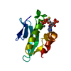
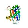
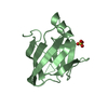
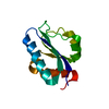
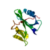
| Title | CRYSTAL STRUCTURE OF IIBCELLOBIOSE FROM ESCHERICHIA COLI | ||||||
 Components Components | ENZYME IIB OF THE CELLOBIOSE-SPECIFIC PHOSPHOTRANSFERASE SYSTEM | ||||||
 Keywords Keywords | PHOSPHOTRANSFERASE / PHOSPHOENOLPYRUVATE DEPENDENT PHOSPHOTRANSFERASE SYSTEM / IIB ENZYMES / CYSTEINE PHOSPHORYLATION | ||||||
| Function / homology |  Function and homology information Function and homology informationprotein-Npi-phosphohistidine-N,N'-diacetylchitobiose phosphotransferase / protein-phosphocysteine-N,N'-diacetylchitobiose phosphotransferase system transporter activity / N,N'-diacetylchitobiose import / protein-phosphocysteine-sugar phosphotransferase activity / protein-N(PI)-phosphohistidine-carbohydrate phosphotransferase activity / phosphoenolpyruvate-dependent sugar phosphotransferase system / transmembrane transporter complex / kinase activity / cytosol Similarity search - Function | ||||||
| Biological species |  | ||||||
| Method |  X-RAY DIFFRACTION / SYNCHROTRON / MIR / Resolution: 1.8 Å X-RAY DIFFRACTION / SYNCHROTRON / MIR / Resolution: 1.8 Å | ||||||
 Authors Authors | Van Montfort, R.L.M. / Pijning, T. / Kalk, K.H. / Reizer, J. / Saier, M.H. / Thunnissen, M.M.G.M. / Robillard, G.T. / Dijkstra, B.W. | ||||||
 Citation Citation | Journal: Structure / Year: 1997 Title: The structure of an energy-coupling protein from bacteria, IIBcellobiose, reveals similarity to eukaryotic protein tyrosine phosphatases. Authors: van Montfort, R.L. / Pijning, T. / Kalk, K.H. / Reizer, J. / Saier Jr., M.H. / Thunnissen, M.M. / Robillard, G.T. / Dijkstra, B.W. | ||||||
| History |
|
- Structure visualization
| Structure viewer | Molecule: MolmilJmol/JSmol |
|---|
- Downloads & links
-Download
| PDBx/mmCIF format | 1iib.cif.gz | 51.4 KB | Display | PDBx/mmCIF format |
|---|---|---|---|---|
| PDB format | pdb1iib.ent.gz | 37.6 KB | Display | PDB format |
| PDBx/mmJSON format | 1iib.json.gz | Tree view | PDBx/mmJSON format | |
| Others |  Other downloads Other downloads |
-Validation report
| Arichive directory | https://data.pdbj.org/pub/pdb/validation_reports/ii/1iibftp://data.pdbj.org/pub/pdb/validation_reports/ii/1iib | HTTPS FTP |
|---|
-Related structure data
| Similar structure data |
|---|


-Links
 PDBj
PDBj- Assembly
| Deposited unit | 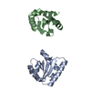
| ||||||||
|---|---|---|---|---|---|---|---|---|---|
| 1 | 
| ||||||||
| 2 | 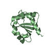
| ||||||||
| Unit cell |
| ||||||||
| Noncrystallographic symmetry (NCS) | NCS oper: (Code: given Matrix: (0.995894, -0.002445, -0.090488), Vector: |
-Components
| #1: Protein | Mass: 11423.449 Da / Num. of mol.: 2 / Fragment: ENZYME IIB / Mutation: C10S Source method: isolated from a genetically manipulated source Source: (gene. exp.) Production host: Strain (production host): W3110 References: UniProt: P69795, protein-Npi-phosphohistidine-sugar phosphotransferase #2: Water | ChemComp-HOH / |  Mass: 18.015 Da / Num. of mol.: 128 / Source method: isolated from a natural source / Formula: H2O Mass: 18.015 Da / Num. of mol.: 128 / Source method: isolated from a natural source / Formula: H2O |
|---|
-Experimental details
-Experiment
| Experiment | Method: X-RAY DIFFRACTION / Number of used crystals: 1 |
|---|
- Sample preparation
| Crystal | Density Matthews: 2.2 Å3/Da / Density % sol: 44 % Description: THE NATIVE DATASET USED WAS OBTAINED BY MERGING OF A 2.6 ANGSTROM IN HOUSE DATA SET WITH A 1.8 ANGSTROM X31 DATASET. THE STATISTICS OF THE X-31 DATASET ARE GIVEN ABOVE. | ||||||||||||||||||||||||||||||||||||||||||||||||||||||||||||||||||
|---|---|---|---|---|---|---|---|---|---|---|---|---|---|---|---|---|---|---|---|---|---|---|---|---|---|---|---|---|---|---|---|---|---|---|---|---|---|---|---|---|---|---|---|---|---|---|---|---|---|---|---|---|---|---|---|---|---|---|---|---|---|---|---|---|---|---|---|
| Crystal grow | pH: 6.5 / Details: pH 6.5 | ||||||||||||||||||||||||||||||||||||||||||||||||||||||||||||||||||
| Crystal grow | *PLUS Temperature: 4 ℃ / Method: vapor diffusion, hanging drop / Details: macroseeding | ||||||||||||||||||||||||||||||||||||||||||||||||||||||||||||||||||
| Components of the solutions | *PLUS
|
-Data collection
| Diffraction | Mean temperature: 293 K |
|---|---|
| Diffraction source | Source: SYNCHROTRON / Site: EMBL/DESY, HAMBURG  / Beamline: X31 / Wavelength: 0.92 / Beamline: X31 / Wavelength: 0.92 |
| Detector | Type: MARRESEARCH / Detector: IMAGE PLATE / Date: May 1, 1995 |
| Radiation | Monochromatic (M) / Laue (L): M / Scattering type: x-ray |
| Radiation wavelength | Wavelength: 0.92 Å / Relative weight: 1 |
| Reflection | Resolution: 1.8→30 Å / Num. obs: 16165 / % possible obs: 82.8 % / Observed criterion σ(I): 3 / Redundancy: 3.7 % / Rmerge(I) obs: 0.061 |
| Reflection shell | Resolution: 1.8→1.83 Å / Rmerge(I) obs: 0.122 / % possible all: 74.1 |
| Reflection | *PLUS Rmerge(I) obs: 0.061 |
| Reflection shell | *PLUS Rmerge(I) obs: 0.122 |
- Processing
| Software |
| ||||||||||||||||||||||||||||||||||||||||||||||||||||||||||||
|---|---|---|---|---|---|---|---|---|---|---|---|---|---|---|---|---|---|---|---|---|---|---|---|---|---|---|---|---|---|---|---|---|---|---|---|---|---|---|---|---|---|---|---|---|---|---|---|---|---|---|---|---|---|---|---|---|---|---|---|---|---|
| Refinement | Method to determine structure: MIR / Resolution: 1.8→10 Å / Isotropic thermal model: RESTRAINED / Cross valid method: THROUGHOUT / Details: NCS-RESTRAINTS RELEASED AT 1.8 ANGSTROM RESOLUTION
| ||||||||||||||||||||||||||||||||||||||||||||||||||||||||||||
| Displacement parameters | Biso mean: 21.9 Å2 | ||||||||||||||||||||||||||||||||||||||||||||||||||||||||||||
| Refine analyze | Luzzati d res low obs: 10 Å / Luzzati sigma a obs: 0.22 Å | ||||||||||||||||||||||||||||||||||||||||||||||||||||||||||||
| Refinement step | Cycle: LAST / Resolution: 1.8→10 Å
| ||||||||||||||||||||||||||||||||||||||||||||||||||||||||||||
| Refine LS restraints |
| ||||||||||||||||||||||||||||||||||||||||||||||||||||||||||||
| Xplor file |
| ||||||||||||||||||||||||||||||||||||||||||||||||||||||||||||
| Software | *PLUS Name: X-PLOR / Version: 3.1 / Classification: refinement | ||||||||||||||||||||||||||||||||||||||||||||||||||||||||||||
| Refinement | *PLUS | ||||||||||||||||||||||||||||||||||||||||||||||||||||||||||||
| Solvent computation | *PLUS | ||||||||||||||||||||||||||||||||||||||||||||||||||||||||||||
| Displacement parameters | *PLUS | ||||||||||||||||||||||||||||||||||||||||||||||||||||||||||||
| Refine LS restraints | *PLUS
|
