 Movie
Movie Controller


+ Open data

- Basic information
| Entry | Database: PDB / ID: 4ix9 | ||||||
|---|---|---|---|---|---|---|---|
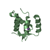
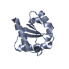
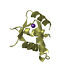
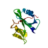
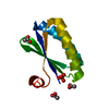
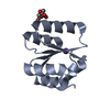
| Title | Crystal structure of subunit F of V-ATPase from S. cerevisiae | ||||||
 Components Components | V-type proton ATPase subunit F | ||||||
 Keywords Keywords | HYDROLASE / V-ATPase / stalk subunit / subunit F / Rossmann fold / Regulatory / coupling | ||||||
| Function / homology |  Function and homology information Function and homology informationInsulin receptor recycling / Transferrin endocytosis and recycling / ROS and RNS production in phagocytes / Golgi lumen acidification / Amino acids regulate mTORC1 / vacuolar proton-transporting V-type ATPase, V1 domain / endosomal lumen acidification / vacuolar acidification / fungal-type vacuole membrane / proton-transporting ATPase activity, rotational mechanism ...Insulin receptor recycling / Transferrin endocytosis and recycling / ROS and RNS production in phagocytes / Golgi lumen acidification / Amino acids regulate mTORC1 / vacuolar proton-transporting V-type ATPase, V1 domain / endosomal lumen acidification / vacuolar acidification / fungal-type vacuole membrane / proton-transporting ATPase activity, rotational mechanism / proton transmembrane transport / Golgi membrane / membrane Similarity search - Function | ||||||
| Biological species |  | ||||||
| Method |  X-RAY DIFFRACTION / SYNCHROTRON / MAD / Resolution: 2.33 Å X-RAY DIFFRACTION / SYNCHROTRON / MAD / Resolution: 2.33 Å | ||||||
 Authors Authors | Basak, S. / Balakrishna, A.M. / Manimekalai, M.S.S. / Gruber, G. | ||||||
 Citation Citation | Journal: J.Biol.Chem. / Year: 2013 Title: Crystal and NMR structures give insights into the role and dynamics of subunit F of the eukaryotic V-ATPase from Saccharomyces cerevisiae Authors: Basak, S. / Lim, J. / Manimekalai, M.S.S. / Balakrishna, A.M. / Gruber, G. | ||||||
| History |
|
- Structure visualization
| Structure viewer | Molecule: MolmilJmol/JSmol |
|---|
- Downloads & links
-Download
| PDBx/mmCIF format | 4ix9.cif.gz | 93.9 KB | Display | PDBx/mmCIF format |
|---|---|---|---|---|
| PDB format | pdb4ix9.ent.gz | 74 KB | Display | PDB format |
| PDBx/mmJSON format | 4ix9.json.gz | Tree view | PDBx/mmJSON format | |
| Others |  Other downloads Other downloads |
-Validation report
| Arichive directory | https://data.pdbj.org/pub/pdb/validation_reports/ix/4ix9ftp://data.pdbj.org/pub/pdb/validation_reports/ix/4ix9 | HTTPS FTP |
|---|
-Related structure data
| Similar structure data |
|---|


-Links
 PDBj
PDBj

- Assembly
| Deposited unit | 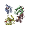
| ||||||||||||||||||||
|---|---|---|---|---|---|---|---|---|---|---|---|---|---|---|---|---|---|---|---|---|---|
| 1 | 
| ||||||||||||||||||||
| 2 | 
| ||||||||||||||||||||
| 3 | 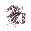
| ||||||||||||||||||||
| 4 | 
| ||||||||||||||||||||
| Unit cell |
| ||||||||||||||||||||
| Components on special symmetry positions |
| ||||||||||||||||||||
| Noncrystallographic symmetry (NCS) | NCS oper:
|
-Components
| #1: Protein | Mass: 10760.775 Da / Num. of mol.: 4 Fragment: Coupling and Regulatory Subunit F, UNP residues 1-94 Mutation: I69M Source method: isolated from a genetically manipulated source Source: (gene. exp.) Strain: ATCC 204508 / S288c / Gene: VMA7, YGR020C / Plasmid: pET9d / Production host:  References: UniProt: P39111, H+-transporting two-sector ATPase #2: Chemical | ChemComp-TRS /   Mass: 122.143 Da / Num. of mol.: 4 / Source method: obtained synthetically / Formula: C4H12NO3 / Comment: pH buffer*YM Mass: 122.143 Da / Num. of mol.: 4 / Source method: obtained synthetically / Formula: C4H12NO3 / Comment: pH buffer*YM#3: Chemical | ChemComp-GOL /   Mass: 92.094 Da / Num. of mol.: 5 / Source method: obtained synthetically / Formula: C3H8O3 Mass: 92.094 Da / Num. of mol.: 5 / Source method: obtained synthetically / Formula: C3H8O3#4: Water | ChemComp-HOH / |  Mass: 18.015 Da / Num. of mol.: 245 / Source method: isolated from a natural source / Formula: H2O Mass: 18.015 Da / Num. of mol.: 245 / Source method: isolated from a natural source / Formula: H2OHas protein modification | Y | |
|---|
-Experimental details
-Experiment
| Experiment | Method: X-RAY DIFFRACTION / Number of used crystals: 1 |
|---|
- Sample preparation
| Crystal | Density Matthews: 2.25 Å3/Da / Density % sol: 45.29 % |
|---|---|
| Crystal grow | Temperature: 291 K / Method: vapor diffusion, hanging drop / pH: 8.8 Details: 30% PEG 4000, 0.05M MgCl2,6H2O, 0.1M Tris HCl pH 8.8, 10% glycerol, VAPOR DIFFUSION, HANGING DROP, temperature 291K |
-Data collection
| Diffraction | Mean temperature: 100 K | ||||||||||||
|---|---|---|---|---|---|---|---|---|---|---|---|---|---|
| Diffraction source | Source: SYNCHROTRON / Site: NSRRC  / Beamline: BL13B1 / Wavelength: 0.978836, 0.978683, 0.963626 / Beamline: BL13B1 / Wavelength: 0.978836, 0.978683, 0.963626 | ||||||||||||
| Detector | Type: ADSC QUANTUM 315 / Detector: CCD / Date: Mar 31, 2012 Details: Vertically Collimating Premirror, Toroidal Focusing Mirror | ||||||||||||
| Radiation | Monochromator: Double Crystal Si(111) Monochromator / Protocol: MAD / Monochromatic (M) / Laue (L): M / Scattering type: x-ray | ||||||||||||
| Radiation wavelength |
| ||||||||||||
| Reflection | Resolution: 2.33→50 Å / Num. all: 17114 / Num. obs: 17018 / % possible obs: 99.5 % / Observed criterion σ(F): 2 / Observed criterion σ(I): 2 / Redundancy: 6.5 % / Biso Wilson estimate: 37.6 Å2 / Rmerge(I) obs: 0.084 / Net I/σ(I): 18.8 | ||||||||||||
| Reflection shell | Resolution: 2.33→2.41 Å / Redundancy: 4.6 % / Rmerge(I) obs: 0.434 / Mean I/σ(I) obs: 2.2 / Num. unique all: 1612 / % possible all: 96.7 |
- Processing
| Software |
| |||||||||||||||||||||||||||||||||||||||||||||
|---|---|---|---|---|---|---|---|---|---|---|---|---|---|---|---|---|---|---|---|---|---|---|---|---|---|---|---|---|---|---|---|---|---|---|---|---|---|---|---|---|---|---|---|---|---|---|
| Refinement | Method to determine structure: MAD / Resolution: 2.33→26.53 Å / Cor.coef. Fo:Fc: 0.966 / Cor.coef. Fo:Fc free: 0.939 / SU B: 6.446 / SU ML: 0.155 / Cross valid method: THROUGHOUT / ESU R: 0.402 / ESU R Free: 0.229 / Stereochemistry target values: MAXIMUM LIKELIHOOD / Details: HYDROGENS HAVE BEEN USED IF PRESENT IN THE INPUT
| |||||||||||||||||||||||||||||||||||||||||||||
| Solvent computation | Ion probe radii: 0.8 Å / Shrinkage radii: 0.8 Å / VDW probe radii: 1.2 Å / Solvent model: MASK | |||||||||||||||||||||||||||||||||||||||||||||
| Displacement parameters | Biso mean: 39.034 Å2
| |||||||||||||||||||||||||||||||||||||||||||||
| Refinement step | Cycle: LAST / Resolution: 2.33→26.53 Å
| |||||||||||||||||||||||||||||||||||||||||||||
| Refine LS restraints |
| |||||||||||||||||||||||||||||||||||||||||||||
| LS refinement shell | Resolution: 2.328→2.389 Å / Total num. of bins used: 20
|
