 Movie
Movie Controller


+ Open data

- Basic information
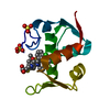
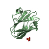
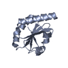
| Entry | Database: PDB / ID: 3v7q | ||||||
|---|---|---|---|---|---|---|---|
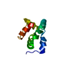
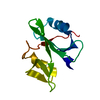
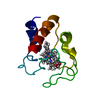
| Title | Crystal structure of B. subtilis YlxQ at 1.55 A resolution | ||||||
 Components Components | Probable ribosomal protein ylxQ | ||||||
 Keywords Keywords | RNA BINDING PROTEIN / L7Ae superfamily / K-turn binding / K-turn RNA / Hypothetical ribosomal protein | ||||||
| Function / homology |  Function and homology information Function and homology information | ||||||
| Biological species |  | ||||||
| Method |  X-RAY DIFFRACTION / SYNCHROTRON / MOLECULAR REPLACEMENT / Resolution: 1.55 Å X-RAY DIFFRACTION / SYNCHROTRON / MOLECULAR REPLACEMENT / Resolution: 1.55 Å | ||||||
 Authors Authors | Baird, N.J. / Zhang, J. / Hamma, T. / Ferre-D'Amare, A.R. | ||||||
 Citation Citation | Journal: Rna / Year: 2012 Title: YbxF and YlxQ are bacterial homologs of L7Ae and bind K-turns but not K-loops. Authors: Baird, N.J. / Zhang, J. / Hamma, T. / Ferre-D'Amare, A.R. | ||||||
| History |
|
- Structure visualization
| Structure viewer | Molecule: MolmilJmol/JSmol |
|---|
- Downloads & links
-Download
| PDBx/mmCIF format | 3v7q.cif.gz | 77.7 KB | Display | PDBx/mmCIF format |
|---|---|---|---|---|
| PDB format | pdb3v7q.ent.gz | 56.8 KB | Display | PDB format |
| PDBx/mmJSON format | 3v7q.json.gz | Tree view | PDBx/mmJSON format | |
| Others |  Other downloads Other downloads |
-Validation report
| Arichive directory | https://data.pdbj.org/pub/pdb/validation_reports/v7/3v7qftp://data.pdbj.org/pub/pdb/validation_reports/v7/3v7q | HTTPS FTP |
|---|
-Related structure data
| Related structure data |  3v7eC  3on1S C: citing same article ( S: Starting model for refinement |
|---|---|
| Similar structure data |


-Links
 PDBj
PDBj


- Assembly
| Deposited unit | 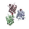
| ||||||||
|---|---|---|---|---|---|---|---|---|---|
| 1 | 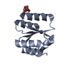
| ||||||||
| 2 | 
| ||||||||
| 3 | 
| ||||||||
| Unit cell |
|
-Components
| #1: Protein | Mass: 11128.995 Da / Num. of mol.: 3 Source method: isolated from a genetically manipulated source Source: (gene. exp.) #2: Chemical |   Mass: 192.124 Da / Num. of mol.: 3 / Source method: obtained synthetically / Formula: C6H8O7 Mass: 192.124 Da / Num. of mol.: 3 / Source method: obtained synthetically / Formula: C6H8O7#3: Chemical |   Mass: 39.098 Da / Num. of mol.: 2 / Source method: obtained synthetically / Formula: K Mass: 39.098 Da / Num. of mol.: 2 / Source method: obtained synthetically / Formula: K#4: Water | ChemComp-HOH / |  Mass: 18.015 Da / Num. of mol.: 246 / Source method: isolated from a natural source / Formula: H2O Mass: 18.015 Da / Num. of mol.: 246 / Source method: isolated from a natural source / Formula: H2O |
|---|
-Experimental details
-Experiment
| Experiment | Method: X-RAY DIFFRACTION / Number of used crystals: 1 |
|---|
- Sample preparation
| Crystal | Density Matthews: 2.18 Å3/Da / Density % sol: 43.53 % |
|---|---|
| Crystal grow | Temperature: 277 K / Method: vapor diffusion, sitting drop / pH: 7 Details: 0.2 M ammonium acetate pH 7.0, 0.1 M sodium citrate pH 5.6, and 30% (w/v) PEG 4000, VAPOR DIFFUSION, SITTING DROP, temperature 277K |
-Data collection
| Diffraction | Mean temperature: 298 K |
|---|---|
| Diffraction source | Source: SYNCHROTRON / Site: ALS  / Beamline: 5.0.1 / Wavelength: 1 Å / Beamline: 5.0.1 / Wavelength: 1 Å |
| Detector | Type: ADSC QUANTUM 210r / Detector: CCD / Date: May 10, 2010 |
| Radiation | Monochromator: Asymmetric curved crystal of Si(220) / Protocol: SINGLE WAVELENGTH / Monochromatic (M) / Laue (L): M / Scattering type: x-ray |
| Radiation wavelength | Wavelength: 1 Å / Relative weight: 1 |
| Reflection | Resolution: 1.5→30 Å / Num. all: 42151 / Num. obs: 41302 / % possible obs: 98 % / Observed criterion σ(F): 0 / Observed criterion σ(I): -3 / Biso Wilson estimate: 17.6 Å2 |
| Reflection shell | Resolution: 1.55→1.61 Å / Redundancy: 5.5 % / Num. unique all: 4152 / % possible all: 98.4 |
- Processing
| Software |
| ||||||||||||||||||||||||||||||||||||
|---|---|---|---|---|---|---|---|---|---|---|---|---|---|---|---|---|---|---|---|---|---|---|---|---|---|---|---|---|---|---|---|---|---|---|---|---|---|
| Refinement | Method to determine structure: MOLECULAR REPLACEMENT Starting model: 3on1 Resolution: 1.55→19.97 Å / Rfactor Rfree error: 0.004 / Data cutoff high absF: 149294.2 / Data cutoff low absF: 0 / Isotropic thermal model: RESTRAINED / Cross valid method: THROUGHOUT / σ(F): 0 / σ(I): 0 / Stereochemistry target values: Engh & Huber / Details: BULK SOLVENT MODEL USED
| ||||||||||||||||||||||||||||||||||||
| Solvent computation | Solvent model: FLAT MODEL / Bsol: 50.8237 Å2 / ksol: 0.4 e/Å3 | ||||||||||||||||||||||||||||||||||||
| Displacement parameters | Biso mean: 21.3 Å2
| ||||||||||||||||||||||||||||||||||||
| Refine analyze |
| ||||||||||||||||||||||||||||||||||||
| Refinement step | Cycle: LAST / Resolution: 1.55→19.97 Å
| ||||||||||||||||||||||||||||||||||||
| Refine LS restraints |
| ||||||||||||||||||||||||||||||||||||
| Refine LS restraints NCS | NCS model details: NONE | ||||||||||||||||||||||||||||||||||||
| LS refinement shell | Resolution: 1.55→1.56 Å / Rfactor Rfree error: 0.011 / Total num. of bins used: 6
| ||||||||||||||||||||||||||||||||||||
| Xplor file |
|
